Knee arthroscopy may help with the excruciating symptoms of a variety of conditions that harm the soft tissues and cartilage surfaces around the joint. Meniscus removal (partial meniscectomy), meniscus repair, and meniscus transplanting are among the common arthroscopic operations for the knee.
Injury to one or more of the ligaments, tendons, cartilage, bones, or muscles that make up the knee joint constitutes a knee injury. These injuries can be caused by falling, twisting the knee violently, heavy impact from a car accident, or other forces.
A 55 year-old patient visited our office with complaints regarding right knee pain. She has been limping. She tried exercises and stretching and NSAIDs but has not helped.
For associated symptoms, patient reported buckling but reports no weakness, no numbness, no tingling, no swelling, no redness, no warmth, no ecchymosis, no catching/locking, no popping/clicking, no grinding, no instability, no radiation, no drainage, no fever, no chills, no weight loss, no change in bowel/bladder habits, and no tenderness.
For quality, she reported aching and burning. For severity, she reported moderate. For context, twisting. For alleviating factors: ice, rest, and limited weight bearing.
The patient presented an MRI result that showed undersurface tear of the medial meniscal body resulting in a small peripheral meniscal flap. Small radial tear at the free edge of the lateral meniscal body. Mild to moderate cartilage loss in the medial compartment and a small joint effusion.
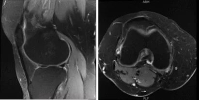
MRI-3T Knee non-contrast
We discussed the treatment options for the patient’s diagnosis, which included living with the extremity as it is, organized exercises, medicines, injections and surgical options. We also discussed the nature and purpose of the treatment options along with the expected risks and benefits.
I educated the patient regarding the inherent and unavoidable risks which include, but are not limited to anesthesia, infection, damage to nerves and blood vessels, blood loss, blood clots, and even death were discussed at length.
We also talked about the possibility of not being able to return to prior activities or employment, the need for future surgery, and complex regional pain syndrome. The patient also understands there is a long rehabilitative process that typically follows the surgical procedure.
We talked about the possibility of not being able to alleviate all of the discomfort. Also, I explained there is no guarantee all the function and strength will return. The patient also understands the risks of re-tear or failure to heal. The patient understands implants may be utilized during this surgery.
The patient expressed understanding of these risks and has elected to proceed with surgery. We have discussed the surgical procedure as well as the realistic expectations regarding the risks, outcome and post operative protocol.
The patient was taken to the operating room where she was placed on a well-padded operating room table. Preop antibiotic was given. General anesthesia was induced. The right lower extremity was prepped and draped aseptically in the usual fashion. The tourniquet was elevated to 275 mmHg.
A lateral arthroscopic entry portal was made. The arthroscope was entered. The Medical entry portal was made with the use of a spinal needle. Examination of the medial tibiofemoral compartment showed flap tear of the posterior horn as well as body of the medial meniscus.
Debridement was done with the use of biter as well as shavers. The meniscus was debrided to a balanced margin. There was an interior flap of the margin of the periphery of the body of the meniscus also. This was also removed with the use of shavers.
There was a grade 1 to grade 2 osteochondral lesion of the medial femoral condyle which was debrided with use of shaver and ablator. Examination of the intercondylar notch showed degenerative ACL but intact.
Examination of the lateral tibiofemoral compartment showed intact femoral cartilage. There was fraying and tear of the medial margin of the lateral meniscus which was debrided with the use of shaver and biters.
Examination of the patellofemoral compartment showed grade 1 to grade 2 tear of the lateral facet of the patella which was debrided again with the shaver. There was no osteochondral lesion on the trochlea. The final picture was taken and saved.
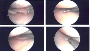
Intraoperative Arthroscopy Images
The knee was thoroughly irrigated and drained. Closure was done with the use of # 3-0 nylon. Then, 9 cc of 0.5% Naropin mixed with 40 mg of Depo-Medrol was injected into the knee.
Dressing was done with the use of Adaptic, 4 x 4, ABD, Webril, and Ace wrap. The patient was extubated in recovery in stable condition.
The patient was seen for post operative check up. We have decided to do formal physical therapy as well as a home exercise program for rehabilitation of the knee.
The patient did well after the surgery and continued physical therapy. Patient checked in for a follow up visit after a month and seen significant improvement on her knee.
Disclaimer – Patient’s name, age, sex, dates, events have been changed or modified to protect patient privacy.
The content on this page has been authored, edited, or approved by the doctors below, and was last reviewed for accuracy on October 19, 2025.
