Accidents like falls or sports injuries are often to blame for common injuries like wrist sprains. Rest, ice, compression, and over-the-counter medication are the standard home treatments for a sprained wrist.
But sometimes, injuries like this require attention from a physician. The surgery usually performed to an injured wrist is called Open Reduction Internal Fixation (ORIF) that helps to restore the articular surface of the radiocarpal joint while respecting soft tissue integrity.
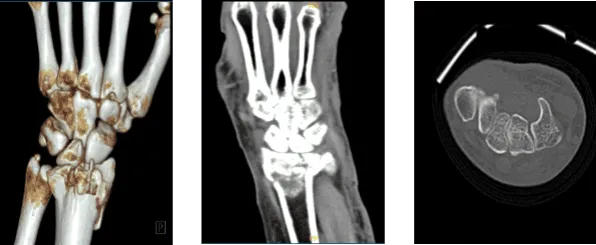
The patient presented today is 68-year-old, female and having a complaint of her left hand and wrist after getting injured because of a fall. Her Xray result showed a fracture but we wanted to make sure of the diagnosis. We agreed to take CT-Scan.
We discussed risks and benefits including infection, bleeding, injury to adjacent nerves and vessels, rehabilitation, failure and need for repeat surgery, continuation of splint, systemic complications including blood clots cardiac or pulmonary, neurological complications. The patient understood and signed an informed consent.
The patient was taken to the operating room where she was placed on well-padded operating table. General anesthesia was induced. Left upper extremity was put in a tourniquet and prepped and draped aseptically in the usual fashion.
Preoperative antibiotics were given. Esmarch was used to exsanguinate the left upper extremity. Timeout was called.
Closed reduction was done and a K-wire was inserted to hold the reduction in place. an incision was given over the tendon of the flexor carpi radialis. The adhered sheath of the flexor carpi radialis was cut and The tendon was retracted medially.
The posterior sheath was also cut in the line of incision. was cut with the use of Bovie along the radial border. The fracture could be seen. The fracture was now reduced in multiple fragments. The K-wire was removed and a final reduction of the fracture was again done with manipulation.
A plate was put on the fracture volarly and held with Olive K-wires. The plate was checked. The plate was fixed proximally with the use of a cortical screw. Pictures were taken and found to be satisfactory.
The distal fragment was brought onto the bed with the use of a nonlocking screw. Final fixation of the plate to the bone was done with the use of locking screws distally and a combination of locking and nonlocking screws proximally.
Two screws were exchanged to a shorter screw of appropriate length. Final pictures were taken and saved. The wound was thoroughly irrigated. Tourniquet was released. Hemostasis was then achieved.
Incision was closed in layers using #2-0 Vicryl and #3-0 Monocryl. Dressing was done with the use of Xeroform and 4×8. Webril and long-arm splint. The patient was extubated in the recovery in a stable condition.
Patient returned to our office after a week of operation, she had X Ray results with her. Post Operative Exam: General Appearance: swelling, tenderness, and erythema and wound clean and dry, no warmth, appropriate range of motion, and neurovascular intact.
X rays were reviewed and discussed. We agreed to go with conservative management for now. Patients take Benadryl for itching and long splints are applied. Follow up after three weeks.
Four-weeks post operative checkup. She has loosened her splint and has been scratching under the ace wraps. Post Operative Exam: General Appearance: no swelling, tenderness, or warmth and wound clean and dry, appropriate range of motion, and neurovascular intact. Long arm splint was removed and wrist brace applied and discussed at home PT program.
Eight-weeks post-operative checkup. She has been using a brace. She has been using it and her pain has improved. Wrist brace to be weaned off and continuous of home PT program.
Patient got well and tolerated the surgery. With the continuous follow up visit, continuous PT, the patient is able to recover and get back to her normal routine.
Disclaimer – Patient’s name, age, sex, dates, events have been changed or modified to protect patient privacy.
The content on this page has been authored, edited, or approved by the doctors below, and was last reviewed for accuracy on October 19, 2025.
