Case Study: Management of a 35-year-old Female
with a Tear of the Long Head of Biceps
The patient presents to our office with complaints of left r of the long head of the biceps te and weakness for the past 2 months. She reportedly had a slip and fall on an outstretched hand. She reported bruising and swelling in front of the upper arm immediately following the injury. She did not seek medical attention at the time of the injury and managed the pain with NSAIDs and icing.
The pain in the left shoulder did not subside and she complains of dull vague pain in the shoulder associated with popping and clicking sensation. She complains of sharp pain in front of the upper left arm. The pain gets significantly worse on activities such as lifting, twisting, and bending of the left arm.
The patient is a former smoker (quit smoking five years ago). She denies the use of any recreational drugs. She is currently not on any medications and denies any known drug allergies.
On physical examination of the left upper extremity, a swelling was present on the upper part of the left arm (proximal and middle biceps with increased size with the flexing of the elbow). Tenderness was present at the glenohumeral joint region (over biceps tendon). Speed’s and Yergason’s tests were positive. The examination of the contralateral limb and the cervical spine was normal.
MRI of the left shoulder and arm revealed non-visualization of biceps tendon within bicipital groove with threadlike intra-articular remnant indicates biceps tendon tear with distal retraction suggesting a tear of the long head of the biceps tendon. Various treatment options were discussed with the patient at length and the patient opted for surgical management.
The risks and benefits and complications of surgery including bleeding, infection, nonhealing, need for repeat surgery, injury to adjacent nerves and vessels among others were discussed at length with the patient. The patient understood and signed the informed consent.
PREOPERATIVE DIAGNOSIS: Traumatic tear of the left proximal long head of biceps.
POSTOPERATIVE DIAGNOSES:
- Tear of the long head of biceps, left shoulder.
- Fraying of the partial tear of the subscapularis muscle of the left shoulder.
- Type 1 SLAP tear of the labrum of the left shoulder.
OPERATION:
- Shoulder arthroscopy and debridement of the left shoulder involving debridement of the subscapularis muscle and glenoid labrum.
- Open biceps tenodesis, subpectoral approach, on the left shoulder using the button and Bio-Tenodesis screw.
DESCRIPTION OF PROCEDURE: The patient was taken to the operating room where she was placed on a well-padded operating room table. General anesthesia was induced. The preop antibiotic was given. The patient was turned on the right lateral position with the left shoulder up where she was held in a beanbag.
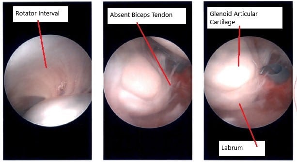
The left upper extremity was prepped and draped aseptically in the usual fashion. Time-out was called. A posterior entry portal was made for the arthroscope. The scope was entered and the glenohumeral joint was examined. There was an absence of biceps tendon. There was fraying of the subscapularis tendon in the lateral part.
Debridement of the labrum, as well as the subscapularis, was performed using the shaver. There was no rotator cuff tear. There was no chondral damage. The scope was entered from the subacromial area, which was found to be normal.
shoulder pain
The scope was removed. The procedure for the subpectoral approach for biceps tenodesis was done. A 3 cm incision was given over the inferior border of the pectoralis major. With sharp and blunt dissection, the bone was teased. The biceps tendon could not be found. It was found to be retracted into scar tissue.
The tendon could be released from the scar tissue. There was some scarring of the muscles also. A whipstitch was passed from the musculotendinous junction distally to proximally on the biceps tendon. The excess biceps tendon was removed.
The suture #2 FiberWire was tied underneath on to itself. The button was put on the suture. Now, the 2.4-mm guidewire was passed into the bicipital groove just under the subpectoral tendon.
Once it was bicortical, unicortical reaming with a 7-mm reamer was performed. The guidewire was removed. Reamer was removed. The button was loaded on its introducer and introduced bicortical. Once it was across, the button was deployed. One of the suture ends was passed through the tendon and tied onto itself.
Now, a 7-mm Bio-Tenodesis EndoButton was used to augment the fixation and the button was flush with the cortex. Finding it in an acceptable position, the wound was thoroughly washed and irrigated.
The closure was done in layers using #0 Vicryl, #2-0 Vicryl, and #3-0 Monocryl. The arthroscopic wounds were closed with #3-0 nylon. Local anesthesia, 15 mL of 1% lidocaine, and 0.5% Marcaine were infiltrated. The dressing was done using 4 x 4, ABD, and tape.
A shoulder immobilizer was applied. The patient will be on Keflex 500 q. 6 for five days and Norco 5/300 mg one to two pills every 4 to 6 hours for pain. The patient is to use ice and elevation.
Disclaimer – Patient’s name, age, sex, dates, events have been changed or modified to protect patient privacy.
My name is Dr. Suhirad Khokhar, and am an orthopaedic surgeon. I completed my MBBS (Bachelor of Medicine & Bachelor of Surgery) at Govt. Medical College, Patiala, India.
I specialize in musculoskeletal disorders and their management, and have personally approved of and written this content.
My profile page has all of my educational information, work experience, and all the pages on this site that I've contributed to.


