Case Study: Elbow Bursectomy: Right Olecranon
in a 74 year-old female
A day case technique is typically used to remove an olecranon bursa. To remove the bursa, a single 5–6 cm-long incision is performed over the prominence of the elbow. Steri-strips and dissolving sutures are used to seal the wound. Olecranon bursitis typically doesn’t require surgical intervention.
A bursectomy may be necessary in severe cases that are resistant to conservative treatment. In either aseptic or septic instances, endoscopic olecranon bursectomy is a successful substitute for open bursectomy if surgical intervention is necessary.
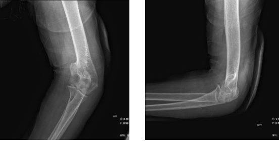
A 74 year-old female patient is here with X-rays of her left elbow and complains the pain from her elbow. According to her X-ray she has Olecranon TBW with increased temperature and redness. However, we discussed the Risks, benefits and alternatives at length.
We advised the patient to take medications and treatment such as Keflex and Mobic for infections, and also, we told her to take rest, use ice for pain and inflammation. And she was told and understood that she may need aspiration/surgery if the pain is not getting better.
The patient will be back in 3 days for her follow up checkup. After three days, the patient went back to the office with the same complaints but the possible infection had healed now. The patient had a bump which had been there for many months.
After the X-Ray was presented and reviewed, we started to discuss treatment options and the patient opted for surgical management. We discussed risks and benefits including injury to adjacent nerves and vessels, infection, bleeding, recurrence, and need for repeat surgery among others.
We also discussed systemic complications. The patient understood and signed an informed consent.
Left elbow X-ray
The patient was taken to the operating room where general anesthesia was induced. Preop antibiotic was given. The right upper extremity was prepped and draped aseptically in usual fashion after application of tourniquet. Time-out was called.
A curvilinear incision going to the radial side was given over the bursa. With the help of the sharp and blunt dissection, the bursa was delineated and excised completely. There was some rupture of the bursa which showed white griseous material draining out.
Culture, sensitivity as well as histopathology was sent. Hemostasis was achieved after removal of the tourniquet. The wound was thoroughly irrigated.
Closure was done using # 3-0 Vicryl and #4-0 nylon. Dressing was done using Adaptic, 4 x 8, Webril, and Ace wrap. A short arm splint was applied. The patient was extubated and moved to Recovery in stable condition.
After one week the patient visits the office for a post operative appointment, no X-rays were indicated. Patients have no other symptoms however; the patient’s biopsy shows Gouty Tophi, S/R is done, Activities as tolerated.
The patient’s Follow Up checkup after 3 weeks. The patient’s progress and development have been undeniable with continued physical treatment and regular attendance at his follow-up checkups.
Disclaimer – Patient’s name, age, sex, dates, events have been changed or modified to protect patient privacy.
I am Vedant Vaksha, Fellowship trained Spine, Sports and Arthroscopic Surgeon at Complete Orthopedics. I take care of patients with ailments of the neck, back, shoulder, knee, elbow and ankle. I personally approve this content and have written most of it myself.
Please take a look at my profile page and don't hesitate to come in and talk.


