Case Study: Arthroscopic Debridement, Decompression
Acromioplasty and Distal Clavicle Excision of the Left Shoulder
The patient is here today and has been complaining of right shoulder pain. Patient states he cannot extend his arm. The pain is severe in intensity. Patient describes the pain as stabbing. The pain is intermittent, and does disturb sleep.
The pain is associated with swelling but not associated with bruising, tingling, numbness, radiating pain, weakness, bowel or bladder abnormality, gait problem, giving way, or limping, hand function difficulty.
The problem has been getting worse since it started. Lifting makes the symptoms worse. Ice makes the symptoms better.
We did an MRI, which shows rotator cuff tear with biceps tear as well as acromioclavicular arthritis. We gave a cortisone injection which failed. The patient opted for surgical management.
We discussed risks and benefits including bleeding, infection, nonhealing, repeat surgery, chronic shoulder pain, need for rehabilitation, and injury to adjacent nerves and vessels among others. The patient understood and signed an informed consent.
The patient was taken to the operating room where he was placed on a well-padded operating room table. General anesthesia was Induced. He was turned into the left lateral position with the right shoulder up. The right shoulder was prepped and draped aseptically in the usual fashion.
He was held in a bean bag with axillary rolls in a well-padded position. All the bony prominences were well padded. Preoperative antibiotic was given. Aseptic cleaning and prepping and draping were performed in the usual fashion. Time-out was called.
Posterior entry portal for the shoulder was made through the soft spot. Scope was entered into the shoulder glenohumeral joint. Anterior entry portal was made by using a spinal needle and a small incision was given right by the coracoid process. Shaver was introduced from the anterior portal.
Examination of the shoulder showed fraying of the labrum as well as tear of the biceps at its insertion into the labrum. Biceps tenotomy was performed. Rest of the examination was normal. There was no particular site rotator cuff tear.
The scope was entered into the subacromial space where subacromial bursitis was present. Shaving was performed. Shaver was used to debride the subacromial bursa. A lateral anterior portal was made.
The scope was entered from the lateral anterior portal and further shaving was performed on the posterior portal. Acromioplasty was performed for type 2 acromion by using the Bovie followed by a burr. After a thorough acromioplasty was performed, the AC joint was examined and found to have arthritis.
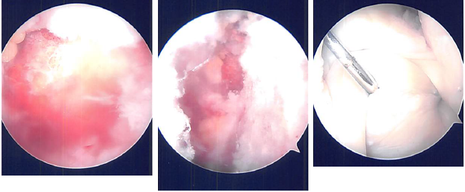
Distal clavicle excision was started from the posterior portal and then the shaver was moved into the anterior portal and distal clavicle by about 1 cm was excised. Pictures were taken and saved.
There was a partial rotator cuff cleared of the bursal side. A thorough examination was performed and it was found to be partial only. There was no deep connection into the joint. So, a decision was done not to repair the rotator cuff tear.
Debridement of the rotator cuff was performed. Final pictures were taken and saved. The shoulder was irrigated and drained. Now, the shoulder was removed from the traction and put in abduction and external rotation. A 3 cm incision was given over the insertion of pectoralis major into the humerus.
Dissection was taken deep by cutting the superficial and deep fascia. The pectoralis major was seen. It was retracted laterally and Hohmann’s retractor was inserted on to the humerus. The biceps tendon was found and brought out using a right angle. A whipstitch was done into the bicep’s tendon.
A hole was drilled finding the biceps tendon in good tension. The whipstitch was done using Ethibond 2 and 3.5. A 4.5 mm cannulated screw with a washer was used to do the biceps tenodesis on the humerus. Excess of tendon was cut. Final pictures were also taken.
The wound was thoroughly washed and disclosed in layer using #0 Vicryl, #2-0 Vicryl and Monocryl. Dressing was performed. Steri-strips were put. The shoulder scope incisions were closed using nylon #3-0. The wound was thoroughly cleaned and dressing was performed using 4×4, ABD and tape.

Intraoperative Arthroscopic Images
After a week post-operative, a patient came into the office to discuss the treatment options. We have decided to proceed with formal physical therapy as well as a home exercise program for the rehabilitation of her shoulder. Stitches were removed.
Patient will continue the ice and elevation for it helps with her condition. Patients regularly followed an office visit every 3-4 weeks. Patient did well after the surgery and continued physical therapy.
Disclaimer – Patient’s name, age, sex, dates, events have been changed or modified to protect patient privacy.

Dr. Vedant Vaksha
I am Vedant Vaksha, Fellowship trained Spine, Sports and Arthroscopic Surgeon at Complete Orthopedics. I take care of patients with ailments of the neck, back, shoulder, knee, elbow and ankle. I personally approve this content and have written most of it myself.
Please take a look at my profile page and don't hesitate to come in and talk.
