The parts of a shattered bone are surgically placed using an open reduction and internal fixation. The shattered bone is held together by rods, screws, plates, sutures, or other devices.
In order to stabilize the segments without dragging the bone to the plate, screws are tightened until they “lock” to the threaded screw holes of the plate. Screw insertion cannot change the reduction thanks to locking screws. Nonlocking plate/screw systems necessitate exact plate adaptation to the underlying bone.
A 64 year-old patient was in our office with complaints regarding wrist pain. She was involved in a motor vehicle accident. She has an injury to face and nose also. She has a history of radial head excision many years back.
For aggravating factors, patients report lifting, twisting, pushing/pulling, throwing, weight bearing, and exercise. For associated symptoms, she reports weakness, numbness, and tingling but reports no swelling, no redness, no warmth, no ecchymosis, no catching/locking, no popping/clicking, no buckling, no grinding, no instability, no radiation, no drainage, no fever, no chills, no weight loss, no change in bowel/bladder habits, and no tenderness.
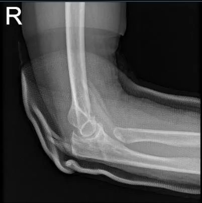
She brought X Ray results for her right elbow with her and has shown prior resection of the right radial head. Mild elbow osteoarthritic degenerative changes. For her right wrist Xray, it showed comminuted angulated distal radial shaft fracture.

Right elbow x-ray
Right wrist x-ray 3 or more views
We discussed the treatment options for the patient’s diagnosis, which included living with the extremity as it is, organized exercises, medicines, injections, and surgical options. We also discussed the nature and purpose of the treatment options along with the expected risks and benefits.
I educated the patient regarding the inherent and unavoidable risks which include, but are not limited to anesthesia, infection, damage to nerves and blood vessels, blood loss, blood clots, and even death were discussed at length.
We also talked about the possibility of not being able to return to prior activities or employment, the need for future surgery, and complex regional pain syndrome. The patient also understands there is a long rehabilitative process that typically follows the surgical procedure.
We talked about the possibility of not being able to alleviate all of the discomfort. Also, I explained there is no guarantee all the function and strength will return. The patient also understands the risks of re-tear or failure to heal. The patient understands implants may be utilized during this surgery.
The patient expressed understanding of these risks and has elected to proceed with surgery. Ample time was given for questions, of which many were addressed. We have discussed the surgical procedure as well as the realistic expectations regarding the risks, outcome, and post operative protocol.
The patient was seen in the operating room where she was placed on a well-padded operating room table. General anesthesia was induced. The right upper extremity was prepped and draped aseptically in the usual fashion. Tourniquet was applied. Esmarch was applied. Tourniquet was elevated.
The volar incision along the tendon of the flexor carpi radialis was made over the distal third of the forearm. The tendon of the flexor carpi radialis was reached with blunt dissection. Hemostasis was achieved.
The anterior sheath of the flexor carpi radialis was incised along the line of incision and retracted medially. The posterior sheath was incised in the line of incision. Pronator quadratus was reached, which was cut with the cautery on the later border of the radius.
The fracture as well as distal radius could be exposed. The fracture was healing in a non-acceptable position. The fracture was mobilized and clean, curette was used. The fracture site was thoroughly washed and irrigated. The fracture was reduced and held with clamps.
An 8-hole distal radius plate was opened and put and found to be in acceptable position. The plate was fixed to the distal radius, proximal and distal to the fracture site with the use of a combination of locking and nonlocking screws. The wound was thoroughly washed.
The wound was closed in layers using #0 Vicryl, #2-0 Vicryl, #3-0 and #4-0 Monocryl. Dressing was applied with the use of Adaptic, 4x8s, Webril. Long-arm splint was applied. The patient was moved to recover in a stable condition. The patient was moving her fingers well. Brachial block was given in the post op period for pain control.
We have decided to proceed with formal physical therapy as well as a home exercise program for rehabilitation of the wrist. The patient did well after the surgery and continued physical therapy. The patient checked in for a follow up visit after a month and saw significant improvement in his wrist.
Disclaimer – Patient’s name, age, sex, dates, events have been changed or modified to protect patient privacy.
The content on this page has been authored, edited, or approved by the doctors below, and was last reviewed for accuracy on October 19, 2025.
