A damaged bone can be stabilized and healed surgically using open reduction and internal fixation (ORIF). This treatment may be required to repair a shattered femur. The big bone in the top of your leg is called the femur.
This bone can become damaged by various traumas, which can result in it breaking into two or more pieces. The proximal phalanx (PP) base intra-articular fractures typically result from an abduction force, which is most frequently seen in sports accidents or falls.
Because collateral ligament avulsion enhances fracture displacement with MP flexion, displaced fractures may not be reducible conservatively. Avulsion of the extensor tendon, commonly known as mallet fractures, or of the articular surface of the distal phalanx can cause intra-articular fractures of the distal phalanx.
A 22-year-old patient male seen in the office with a letter of consent for the surgery. After the letter was verified the patient was taken to the operating room where monitored anesthesia was induced and local anesthesia was injected and a time out was performed. Antibiotics were given. The hand was prepped and draped in sterile fashion.
At this point, traction was put on the finger and small incisions were made for the point to point clamps which were placed to reduce the fracture. After the clamps were placed the fracture was reduced under AP and lateral views.
I then placed two separate .045 Kirschner wires perpendicular to the fracture and to the joint. Then placed an additional .035″ Kirschner wire on the proximal aspect of this long opening fracture fragment, perpendicular to the fracture site.
Then we placed an oblique Kirschher wire on the ulnar side of the proximal phalanx crossing the fracture as well. All of these were confirmed under AP and lateral planes and Kirschner wires were cut under the skin. Splint was placed. The patient was awoken from monitored anesthesia care.
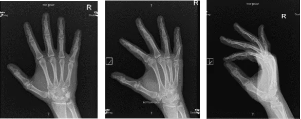
X-ray Right hand minimum 3 years
After one week from the surgery, the patient was discharged. He did well after the surgery and the doctor advised him that he can visit the office as he needed.
Disclaimer – Patient’s name, age, sex, dates, events have been changed or modified to protect patient privacy.
The content on this page has been authored, edited, or approved by the doctors below, and was last reviewed for accuracy on October 19, 2025.
