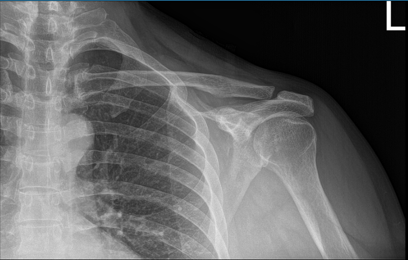
Case Study: Rotator Cuff Repair Performed
to a 66 year-old male patient involved in MVA
The following are the most frequent signs of a rotator cuff injury: Pain while awake and asleep, especially if lying on the injured shoulder. when performing specific actions or raising and lowering your arm, discomfort. having difficulty rotating or elevating your arm.
You must see a doctor if you have any of the following symptoms: It hurts and is difficult to elevate your arm. Popping or clicking sounds or feelings are produced when you move your arm. soreness in the shoulder that worsens at night or whenever you rest your arm.
The patient presented today is a 66-year-old, male, and involved in a motor vehicle accident. He complained today about his bilateral shoulders, but the most painful one is his left shoulder. Patient had no shoulder pain prior to the car accident.
He has been doing therapy and it helped marginally. We suspected that the patient had a rotator cuff tear, however his Xray showed normal radiographs. So, we highly recommended that we take an MRI.
His MRI showed clear mild acromioclavicular hypertrophic change. MRI also showed rotator cuff tear of the supraspinatus along with a tear of the long head of biceps.

MRI of left shoulder
We discussed treatment options and opted for surgical management. We discussed risks and benefits including infection, bleeding, injury to adjacent nerves and vessels, need for shoulder rehabilitation, shoulder stiffness, need for repeat surgery, failure among others.
We also discussed systemic complications including blood clot, cardiac, pulmonary, neurological complications including death. The patient understood and signed an informed consent.
The patient was taken to the operating room where general anesthesia was induced. The patient was given preoperative brachial block. The patient was put in the right lateral position with the left shoulder up. Preoperative antibiotics was given. Left shoulder was prepped and draped aseptically in the usual fashion.
A posterior entry portal was made in the soft spot. Arthroscope was entered. Biceps could be seen torn at the attachment of the glenoid labrum. An anterosuperior portal was made with the use of a spinal needle.
Punch was introduced and the biceps tendon was cut at the junction of the glenoid labrum. There was partial tearing of the subscapularis, which was debrided with the use of a shaver. Rotator cuff tear could be seen from the articular site.
Rest of the examination of the glenohumeral cartilage and ligament was intact. Another arthroscope was entered into the subacromial space. Shaver was introduced from the anterosuperior portal and subacromial bursectomy was performed. Acromial spur was present.
Lateral working portal was made followed by acromioplasty with the use of high-speed 6 mm bur. AC arthritis was present and a plan for distal clavicular excision was also made. The rotator cuff tear was seen. The footprints on the humeral head were prepped with the use of bur.
Debridement of the rotator cuff tear was done. A triple-tailed Arthrex anchor was used and inserted into the humeral head. The suture was passed sequentially x6. The suture was tied on to each other. Good rotator cuff repair was achieved.
Now the distal clavicular excision was performed with the use of high-speed bur from the posterior portal followed by anterosuperior portal. About a centimeter of distal clavicle was excised.
Final pictures were taken and saved. The shoulder was thoroughly irrigated and draped. The arm was taken out of the sling in the preparation for the mini open biceps tenodesis.
The patient’s arm was put in abduction and extension. A 3 cm incision was given along the inferior margin of the pectoralis major tendon near the attachment. With sharp dissection, the deltopectoral fascia was cut.
With blunt dissection, the ball in the bicipital groove was reached and the tendon was extracted with the use of right angle forceps. FiberLoop was used to put stitches through the tendon and extra tendon was cut.
Area for insertion of the biceps tendon was prepped with the use of Bovie followed by bicortical drilling of the Beath pin and a unicortical drilling screw. The tendon was passed with the use of button bicortically and the button was stitched.
The sutures was passed through the tendon and tied on to each other. Now, the 7 mm Endo screw was inserted over the tendon and suture was tied again. Satisfactory tenodesis was achieved.
The wound was thoroughly irrigated and drained. Closure was done in layers using #0 Vicryl, # 2-0 Vicryl, and # 3-0 Monocryl. The arthroscopic portals were closed with the use of # 3-0 nylon.
Dressing was done with the use of Xeroform, 4 x 4, Ace, ABD, and tape. Shoulder immobilizer was applied. The patient was extubated and moved to Recovery in a stable condition.
First-week post-operative. General Appearance: no swelling or warmth; tenderness, passive motion limited, and active motion limited; and wound clean and dry and neurovascular intact. He denies fever and chills.
Four-weeks post-operative. We agreed to go with conservative management for now. Physical Therapy to be started and will continue to take OTC anti-inflammatory medication.
Eight-weeks post-operative. Both shoulders are doing fine and he is improving on left shoulder with PT. Sixteenth-weeks post-operative checkup. He is doing all usual activities without any discomfort. Patient tolerated the surgery well. Recover fast with the help of continuous follow up and home physical therapy.
Disclaimer – Patient’s name, age, sex, dates, events have been changed or modified to protect patient privacy.
I am Vedant Vaksha, Fellowship trained Spine, Sports and Arthroscopic Surgeon at Complete Orthopedics. I take care of patients with ailments of the neck, back, shoulder, knee, elbow and ankle. I personally approve this content and have written most of it myself.
Please take a look at my profile page and don't hesitate to come in and talk.

