Case Study: Open Reduction Internal Fixation
Distal Radius in a 27 year-old female
A damaged bone can be stabilized and healed using a procedure called open reduction and internal fixation (ORIF). This operation might be required to treat your broken ankle. The ankle joint is made up of three bones.
These are the talus (a bone in your foot), fibula (the smaller bone in your leg), and tibia (your shinbone). The distal radius is the portion of the radius that connects to the wrist joint.
Sometimes, a fracture in this region is referred to as a “broken wrist.” The most frequent causes of distal radius fractures are direct blows to the wrist or falling onto an outstretched hand.
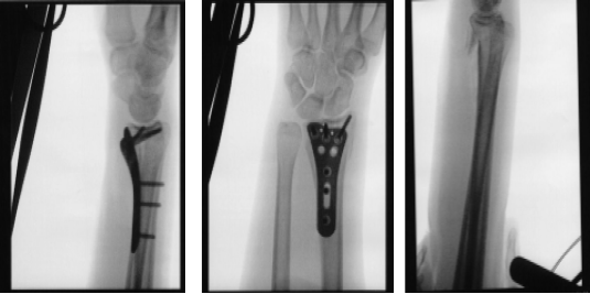
Following a fall, the patient, a 27-year-old female, complains of pain in her right wrist. She has her arm in a brief splint. She underwent a CT scan and x-ray. The distal end of the right side of the radius was found to have a comminuted intra-articular (more than three fragments) fracture after reviewing and discussing the CT and X-ray images.
We spoke about our alternatives for treatment and decided on surgical treatment. Infection, bleeding, damage to nearby nerves and blood vessels, failure to heal, need for additional surgery, wrist pain, rehabilitation, complex regional pain syndrome, systemic complications including blood clots, cardiac, neurological, and pulmonary complications including death were some of the risks and side effects we discussed. Patient gave informed permission after agreeing and signing.

X-ray wrist 3 or more view
The patient was taken to the operating room where she was placed on a well-padded operating table. Supraclavicular block was already given in the holding area. GA was induced. A timeout was called.
Preoperative antibiotics were given. The tourniquet was inflated to 200 mmHg. An incision was given along the volar aspect of the flexor carpi radialis tendon. Hemostasis was achieved.
The anterior sheath of the flexor carpi radialis was incised along the line of incision. The flexor carpi radialis tendon was retracted medially and the posterior sheath was incised along the line of incision. The pronator quadratus was also incised along the line of incision and the bone was reached.
The fracture was healing in malposition. There was not much movement with manual manipulation. An osteotome was used to do osteotomy of the fracture site and reduce it in the correct position.
Once the reduction was achieved, Fluoroscopy showed fracture in acceptable position. The plate was put onto the distal radius and held with olive wires. Finding it in acceptable position on fluoroscopy, the plate was fixed distally by the use of a cortical screw, which was later changed to a locking screw.
Once the plate was in good position, fixation of the proximal fragment with the plate was done by the use of three cortical screws. This was followed by fixation of the distal fragment with the use of locking screws. The central screw was removed and wasted and a locking screw was inserted. The fixation was found to be satisfactory. Final pictures were taken and saved.
The wound was thoroughly irrigated and drained. The tourniquet was deflated. Hemostasis was achieved. Closure was done using 2-0 Vicryl and 3-0 Monocryl. Dressing was done with Xeroform, 4×8, and Webril.
A posterior short arm splint was applied and the hand was in Ace wrap. The patient was extubated. The patient’s arm was put in a shoulder sling. The patient was moved to recovery in a stable condition.
After one week the patient visits the office for her post operative consultation with x-rays of her twist. Her pain is well controlled, she denies fever and chills.
We discussed treatment options including PT, MRI, Injection, surgery and we agreed to go with conservative management for now and the short arm splint is to continue. Ice, Elevation, taking OTC anti-inflammatory meds. The patient did well after the surgery without taking physical therapy.
Disclaimer – Patient’s name, age, sex, dates, events have been changed or modified to protect patient privacy
I am Vedant Vaksha, Fellowship trained Spine, Sports and Arthroscopic Surgeon at Complete Orthopedics. I take care of patients with ailments of the neck, back, shoulder, knee, elbow and ankle. I personally approve this content and have written most of it myself.
Please take a look at my profile page and don't hesitate to come in and talk.

