The clavicle, known by many as the collarbone, is a component of our shoulder. It is a prominent bone that connects the skeleton to the arm. Its main functions are enabling the shoulder to move freely away from the body.
The clavicle, like the rib cage, protects the heart from any external damage. Clavicle is also known as the most fractured bones in the human body. Teenagers and toddlers are more likely than adults to suffer a broken collarbone. After the age of 20, the chance decreases. Then it rises again in older people as they lose bone strength with age.
A 20 year-old male was seen in the office complaining of pain on his left collar bone. He suspected the motor vehicle accident was the reason for it. Pain wakes him up from sleep. It can be very painful and may make it hard to move his arm.
XR was presented in the office and found midshaft clavicular fracture with 1 cm overriding and moderate displacement. Midshaft Clavicle fractures are frequent traumatic injuries produced by a direct impact to the shoulder girdle, and they are most common in young, active adults.
Upon examination of the left shoulder, the patient sits with the scapula protracted and depressed. They are tender to palpation over the anterior aspect of the clavicle. They are non-tender to palpation over the neck, proximal humerus and scapula. There is minor soft tissue swelling and ecchymosis.
The patient has a limited range of motion secondary to discomfort. The patient is unable to tolerate stability testing. They have 5/5 strength at their side, and are neurovascularly intact distally. There are no erythema, warmth or skin lesions present. On examination of the contralateral extremity, the patient is nontender to palpation and has excellent range of motion, stability and strength.
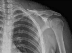
Left Clavicle in AP position
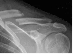
Left Clavicle in axial position
We discussed the treatment options for the patient’s diagnosis, which included: non-surgical and surgical options. We also discussed the nature and purpose of the treatment options along with the expected risks and benefits.
The patient has expressed a desire to proceed with surgery, I educated the patient regarding the inherent and unavoidable risks which include, but are not limited to: anesthesia, infection, damage to nerves and blood vessels, blood loss, blood clots, and even death were discussed at length.
We also talked about the possibility of not being able to return to prior activities or employment, the need for future surgery, and complex regional pain syndrome. The patient also understands there is a long rehabilitative process that typically follows the surgical procedure. Also, I explained there is no guarantee all the function and strength will return.
The patient also understands the risks of failure to heal. The patient understands implants may be utilized during this surgery. The patient acknowledged these risks and decided to continue with surgery.
General anesthesia was induced. IV bag was put in between the two scapulae. The head was elevated by about 30 degrees and the bed was put in reverse Trendelenburg and knees were flexed.
Left upper extremity was prepped and draped aseptically free. Surgical incision was marked and a skin incision was given anterior of the cleft bone of the clavicle.
With sharp and blunt dissection, the bone and the fracture site were reached and exposed. The fracture site was opened up and washed and cleaned with curette. The fracture was reduced and a superior plate was put. The plate was fixed proximally and medially with two screws process for lagging for compression.
Finally, the plate was put and compression was done using a compression screw. Two nonlocking g and one locking screw were used on either side. The final images were taken and found to be satisfactory. Final images were saved.
The wound was washed and closed in layers. A local of 20 mL of 0.5% Marcaine was injected. Closure with Monocryl and Dermabond was done and transparent dressing was applied. The patient’s left upper extremity was put in a shoulder sling.
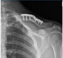
The patient was extubated and moved to the postoperative recovery unit in a stable condition. After two weeks, the patient checked in, with presented XR, and no periprosthetic lucency. The sternoclavicular joint demonstrates normal alignment.
Examination of the left shoulder reveals that the incision is healing well, without evidence of drainage, erythema or warmth. There are small blisters superior and around the incision. Range of motion and strength are progressing appropriately at this stage of rehabilitation.

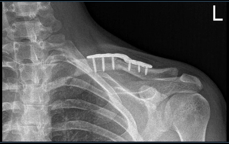
4 weeks X-ray results of left clavicle after surgery
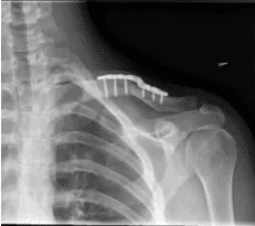
12 weeks X-ray results of left clavicle after surgery
14 weeks X-ray results of left clavicle after surgery
Strength remains 5/5 distally. There is no tenderness at the elbow and wrist. Sensation is intact to light touch distally and there is a brisk capillary refill.
A small non-inflammatory nodule in the axilla. No redness or discharge. With continuous use of shoulder sling and with the help of pendulum exercise the patient recovered well from the surgery.
Disclaimer – Patient’s name, age, sex, dates, events have been changed or modified to protect patient privacy.
The content on this page has been authored, edited, or approved by the doctors below, and was last reviewed for accuracy on October 20, 2025.
