Case Study: Rotator Cuff Repair performed to
Bilateral shoulders to a patient who
consistently experience pain for 4 years
The main cause of shoulder pain in older adults is musculoskeletal disorders that are directly linked to aging, specifically worsening posture. Shoulder discomfort in older adults frequently develops unexpectedly without any involvement in the incident.
According to a study, shoulder injuries including rotator cuff tears most frequently affect elderly persons. In the early 50s, shoulder pain typically first appears.
Patient presents today with bilateral shoulder pain. She is 69 year-old, nothing remembers involving in any form of accident. Patient has undergone physical therapy and still experiences the pain. She explains that both shoulders have been hurting for almost 4 years now.
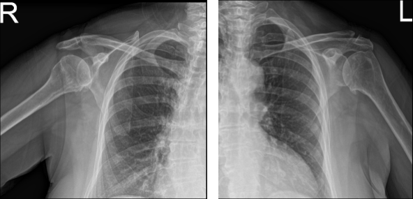
Her pain and restriction of movement are unchanged. She brought her X-ray results today and reviewed them. Her bilateral shoulder showed the impression of mild acromioclavicular and glenohumeral degenerative changes.

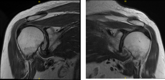
MRI of Left and Right Shoulder
To understand better the condition of her shoulders we decided to take an MRI. Left shoulder MRI impression as follows: Supraspinatus tendinosis with low-grade partial-thickness articular sided tearing. Degeneration of the acromioclavicular and glenohumeral joints.
Truncation of the free edge posterior superior glenoid labrum. Moderate glenohumeral joint effusion. Tendinosis and tenosynovitis of the long of the biceps tendon. Right MRI impression as follows: Degenerative changes of the acromioclavicular and glenohumeral joints. Supraspinatus as with high-grade partial-thickness articular sided tearing. Tear of the posterior superior labrum.

MRI of Left and Right Shoulder
We discussed treatment options and the patient opted for surgical management. We discussed risks and benefits including infection, bleeding, injury to adjunct nerves and blood vessels, shoulder stiffness and rehabilitation, need for repeat surgery, need for shoulder replacement in future.
We also discussed systemic complications including blood clots, cardiac and pulmonary complications including death. The patient understood and signed the informed consent.
The patient was given a supraclavicular block in the holding area. The patient was brought to the operating room where general anesthesia was induced and LMA was inserted. The patient was put in the lateral decubitus position with the left shoulder up.
She was held in a bean bag and all the bony prominences were well padded. A time-out was called. Preoperative antibiotic in the form of clindamycin was given. Surgical incision was marked and incision was given for the posterior and anterior portals. The arthroscope was entered.
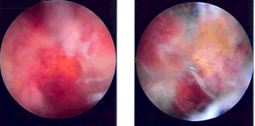
A spinal needle was used to make an anterosuperior portal. Shaver was introduced from the anterosuperior portal. There were grade II to grade III osteoarthritic changes on the glenohumeral joint. There was extensive fraying with a high-grade partial thickness tear of the rotator cuff.
Debridement was done for the glenohumeral joint. After that, the rotator cuff was debrided. The tear was converted into a full thickness tear easily. The biceps were intact. There was synovitis, which was also debrided.
The arthroscope was entered from the anterior portal and the shaver from the posterior portal to complete the debridement. There was a loose body in the glenohumeral joint, which was removed. It seemed to be an osteophyte, which was loose. The arthroscope was entered into the subscapularis region.
The shaver was entered from an anterosuperior portal and subacromial decompression and bursectomy was performed. An accessory lateral portal was made. The scope was entered from the lateral portal and the shaver was entered from the posterior to complete the debridement.
A thermal ablation wand was used to prepare for acromioplasty. A 6.0 burr was used to perform the acromioplasty. After acromioplasty was performed, the rotator cuff tear was debrided and demarcated. The head of the humerus was tapped with the use of shaver and burr for the repair of the rotator cuff.
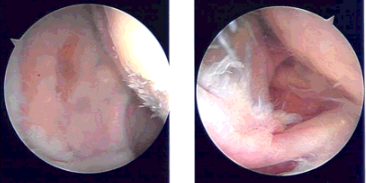
The biceps could be seen. Healicoil triple-loaded was inserted after tapping into the head of the humerus. All six tails of the Healicoil were passed through the rotator cuff tear from anterior to posterior avoiding the biceps. The sutures were tied onto each other with knots. Good repair was achieved.
Pictures were taken. A distal clavicle excision was planned, finding an osteophyte there. A distal clavicle excision was done from the posterior portal following the anterior portal. About a centimeter of distal clavicle was excised. Final pictures were taken and saved.
The shoulder was thoroughly irrigated and drained. Closure was done with the use of 3- 0 nylon. Dressing was done with Xeroform, Webril, ABD, and tape. A shoulder immobilizer was applied. The patient was extubated and moved to recovery in a stable condition.


Intraoperative Images
Patient tolerated the surgery performed on her shoulder. With continuous follow up, OTC anti-inflammatory medications and with the help of physical therapy patient condition is improving every time she visits the office. After 8 weeks, the post-operative patient was able to return to her normal routine.
Disclaimer – Patient’s name, age, sex, dates, events have been changed or modified to protect patient privacy.

Dr. Vedant Vaksha
I am Vedant Vaksha, Fellowship trained Spine, Sports and Arthroscopic Surgeon at Complete Orthopedics. I take care of patients with ailments of the neck, back, shoulder, knee, elbow and ankle. I personally approve this content and have written most of it myself.
Please take a look at my profile page and don't hesitate to come in and talk.
