Someone involved in a car accident has higher possibilities to get a torn meniscus for it’s a very common injury due to any accident. Human knees played an important role in our lives.
It helps us to walk, stand, dance, sports or we can say that every movement that we make, requires the help from our knees. That is mainly the reason why we must pay attention whenever we feel pain in our knees especially when there is an accident involved for it may bring you serious problems.
Patient visits our office today having complaints of his cervical spine and knee pain. He was involved in a motor vehicle accident 3 years ago and suspected maybe the reason why he had pain on his spine and knee.
But his knee makes him uncomfortable. MRIs were advised for us to see the correct diagnosis of his condition. For now, I advised patients to take Flexeril and Mobic for pain medication.
MRI Impression as follows: Oblique tear in the posterior horn of the medial meniscus extending to the inferior surface, peripheral third. Edema in the superolateral aspect of Hoffa’s fat pad suggestive of Hoffa’s fat pad impingement and elevated TT TG distance of 16 mm which can be seen in setting of patellofemoral maltracking and small popliteal cyst.
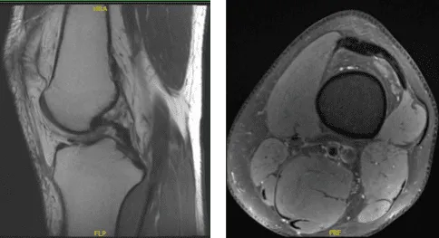
MRI of the back spine
We discussed treatment options and the patient opted for surgical management. We discussed risks of infection, bleeding, failure to heal, knee stiffness, knee pain, and need for knee replacement in the future among others.
We also discussed systemic complications including blood clot, cardiac, pulmonary, and neurological complications including death. The patient understood and signed informed consent.
The patient was taken to the operating room where he was placed on a well-padded operating table. General anesthesia was induced. The left lower extremity was put into a tourniquet and prepped and draped aseptically in the usual fashion.
The left lower extremity was put in a leg holder. The right lower extremity was put in a well-padded pillow. Time-out was called. Preop antibiotics were already given.
Entry portal was made laterally with a lateral parapatellar incision. Arthroscope was entered. Patellofemoral joint showed no injury. A medial plica could be seen. The scope was entered in the medial tibiofemoral compartment. Medial entry portal was made with the use of a spinal needle.
The medial compartment showed no tear of the medial meniscus. There was no cartilage tear. The scope was entered into an intercondylar notch. The ACL was intact with subtle degeneration.
The scope was entered into the lateral compartment where there was some fraying of the medial margin of lateral meniscus which was cleaned with the use of shaver and biter. No other meniscal tear was found.
Scope was entered into the patellofemoral compartment where there was no cartilage damage though there was patellar maltracking. There was a medial synovial plica which was prominent. It was debrided with the use of a shaver.
Final pictures were taken and saved. The knee was irrigated and drained. Knee was closed with nylon # 3-0. Naropin 9 cc mixed with 40 mg of Depo-Medrol was injected into the knee.
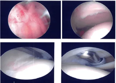
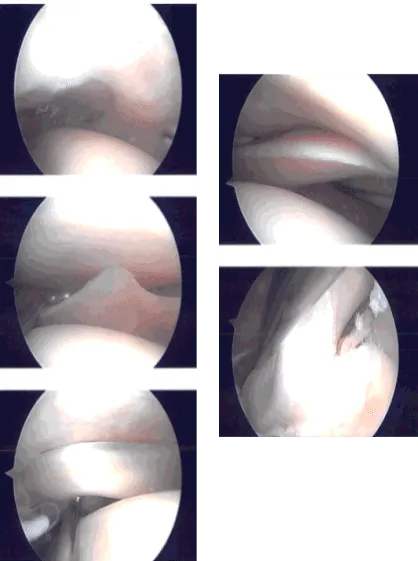
Intraoperative Arthroscopy Images
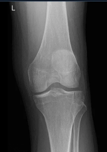
Post Operative Exam: General Appearance: swelling and tenderness and wound clean and dry, no warmth, appropriate range of motion, and neurovascular intact. New X-ray requested to see the improvement of patient condition.
There is a healing fracture of the lateral tibial plateau without depression of the articular surface. The joint spaces are preserved.
Patient got well after surgery; he denied fever and chills. He started physical therapy from the first week of his post operative. He continued to do the R.I.C.E Therapy (Rest/Ice/Compression/Elevation) and abided by the provider’s commendation which really helped him to recover.
Disclaimer – Patient’s name, age, sex, dates, events have been changed or modified to protect patient privacy.