A damaged fragment of cartilage inside the joint is repaired during a meniscus repair procedure on the knee. Common knee ailments include meniscus tears, particularly in sports.
A skilled surgeon can effectively replace damaged knee cartilage with an outpatient surgical technique called arthroscopic meniscus repair. A number of minimally invasive procedures can be used to repair a torn meniscus, but postoperative protection is necessary to allow for recovery.
The patient is a 54 year-old female who had complaints of left knee pain. She has had conservative management, but no benefits. She got an MRI done, which showed a tear of the medial meniscus near the root as well as a degenerative tear of the radial margin of the lateral meniscus.
We discussed treatment options and opted for surgical management. We discussed the risks and benefits including infection, bleeding, injury to adjacent nerves and vessels, retear and the need for repeat surgery, need for rehabilitation, arthritis, need for knee replacement in the future.
We also discussed systemic complications including blood clots, cardiac and pulmonary complications including death. The patient understood and signed the informed consent.
The patient was taken to the operating room where she was placed on a well-padded operating room table. General anesthesia was induced. A time-out was called.
The left lower extremity was prepped and draped aseptically in usual fashion after application of a tourniquet. The tourniquet was inflated to 350 mmHg. A lateral entry portal was made using a transverse incision. The arthroscope was entered.
Examination of the patellofemoral joint showed a grade I to grade II patellofemoral arthritis. Examination of the medial compartment showed a medial meniscus tear near the root. A medical entry portal was made with the use of a spinal needle. The probe was inserted to further examine.
The medial meniscus root was found to be intact. The shaver was used to shave the root with the use of a straight biter and bitters to do a meniscectomy of the medial meniscus.
The osteochondral lesion of the medial femoral condyle was debrided with the use of a shaver. The abrasion chondroplasty was performed over the medial femoral condyle.
Examination of the intercondylar notch showed a degenerative ACL, but the ACL was intact. Examination of the lateral femoral condyle showed a radial margin degenerative tear of the lateral meniscus, which was removed with the use of a shaver.
Examination of the patellofemoral compartment showed an osteochondral lesion, which was debrided with the use of a shaver. Final pictures were taken and saved. The knee was thoroughly irrigated and drained.
Closure was done with the use of 3-0 nylon. 20 cc of Marcaine 0.25% mixed with 40 mg of Depo-Medrol was injected into the knee. Dressing was done with the use of Xeroform, 4×4 gauze, ABD, Webril, and Ace wrap. Tourniquet was deflated. The patient was extubated and moved to recovery in a stable condition.
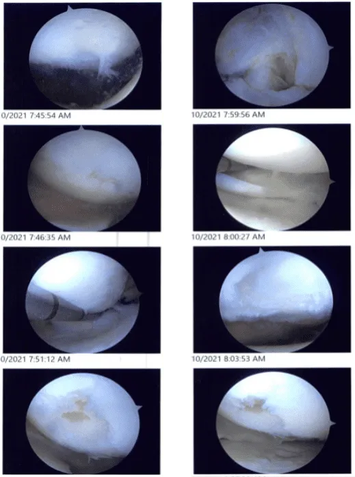
Intraoperative images
After one week the patient was seen in the office for her post operative visits, she had a fractured clavicle right which seems to have healed, she denies fever, chills and she is using a single crutch.
After discussing treatment options, we have decided to proceed with formal physical therapy as well as a home exercise program for rehabilitation of the knee. We went over the arthroscopic pictures and removed the stitches during the visits.
We will continue with ice and elevation of the knee to decrease swelling and pain. We will continue to utilize early mobilization and mechanical prophylaxis to reduce the chances of a deep vein thrombosis.
We will wean them off any narcotic medications and progress to anti-inflammatories and Tylenol as long as there are no contraindications to these medications. We also discussed the risk and benefits and common side effects of taking these medications. I will see them back in three weeks time to evaluate their progress.
After one month the patient visits the office for her follow up checkup, she had a fracture clavicle right which seems to have healed, she denies fever, chills but still using single crutch, however, the patient is started with her physical therapy as well as a home exercise program for rehabilitation of the knee.
After three months the patient seen in the office had a fractured clavicle right which has healed (CT done recently) in malposition. She is able to perform all usual activities. She plans to go back to work.
She denies fever, chills. The patient did well after surgery and her knee is improving. Through regular visits and continued physical therapy, the patient healed and recovered.
Disclaimer – Patient’s name, age, sex, dates, events have been changed or modified to protect patient privacy
The content on this page has been authored, edited, or approved by the doctors below, and was last reviewed for accuracy on October 18, 2025.
