The fracture of the distal radius is one of the most frequent. Either low- or high-energy trauma might be to blame for its occurrence. Conservative treatment is the greatest option for low-energy extra-articular fractures, but ORIF has the highest potential of producing the best results for intra-articular fractures.
Consult a doctor right once if you believe you may have a fractured wrist, especially if you are experiencing numbness, swelling, or difficulty moving your fingers. Poor healing, a reduction in range of motion, and a reduction in grip strength can all result from delayed diagnosis and treatment.
A 71 year-old patient was in our office with complaints of right wrist pain. She stated that she fell at home. She is allergic to codeine and currently not working. She was taken to Brookhaven hospital where the reduction was done, and she was referred to us.
For aggravating factors, the patient reports upstairs and downstairs.
For associated symptoms, she reports pain with motion but reports no weakness, no numbness, no tingling, no swelling, no redness, no warmth, no ecchymosis, no catching/locking, no popping/clicking, no buckling, no grinding, no instability, no radiation, no drainage, no fever, no chills, no weight loss, no change in bowel/bladder habits, and no tenderness.
For location, she reports right. For quality, she reports no change. For severity, she reports severe.
She was here with x-rays of the right wrist which showed 3 views of the right wrist in fiberglass presented for evaluation. Bony detail obscured by overlying fiberglass. Impacted distal transverse distal right radius fracture noted with slight dorsal tilt, post reduction.
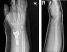
Right wrist x-ray 3 or more views
We discussed treatment options and the patient opted for surgical management. We discussed risks and benefits including infection, bleeding, nonhealing, need for repeat surgery, injury to adjacent nerves and vessels, associated complications among others. The patient understood and signed an informed consent.
The patient was taken to the operating room where she was placed on a well-padded operating room table. General anesthesia was induced. Preoperative antibiotic was given. Right upper extremity was prepped and draped aseptically in a usual fashion.
A volar incision was given along the distal forearm over the flexor carpi radialis tendon. The anterior tendon sheath was reached and cut in line of the incision. The tendon was inspected medially. The posterior was cut along in the line of incision again.
Pronator quadratus was exposed and cut along the radial border with the use of Bovie. The fracture site was opened and washed thoroughly. The fracture was reduced with the help of K-wires. A volar plate was applied. The plate and fracture were found to be in an acceptable position.
The plate was fixed to the distal radius with the use of locking and nonlocking screws. The wound was thoroughly irrigated and draped. Wounds were closed in layers using #2-0 Vicryl and #4-0 Monocryl. Dressing was done using Adaptic, 4×4, and Webril. Short arm splint was applied. The patient was extubated and moved to recover in a stable condition.
We have decided to proceed with formal physical therapy as well as a home exercise program for rehabilitation of the wrist. The patient did well after the surgery and continued physical therapy. The patient checked in for a follow up visit after a month and saw significant improvement in his wrist.
Disclaimer – Patient’s name, age, sex, dates, events have been changed or modified to protect patient privacy.
The content on this page has been authored, edited, or approved by the doctors below, and was last reviewed for accuracy on October 18, 2025.
