Case Study: Arthroscopic Surgery: Shoulder Rotator Cuff
Repair, Medial Meniscectomy with Microfracture
Chondroplasty of the Knee and Shoulder Extensive
Debridement in a 47 year-old female

Reattaching the rotator cuff tendon to the head of the humerus (upper arm bone) is the most common surgical procedure used to treat a torn rotator cuff. In order to create a stable remnant meniscus, unstable flaps of a torn meniscus must be removed.

In order to provide the joint’s gliding surface a smoother surface, two of these procedures are employed to assist smooth out damaged, irregularly shaped cartilage: chondroplasty and microfracture.

Debridement is an arthroscopic operation used to remove debris, damaged cartilage, or tissue from inside a joint, successfully healing the damaged joint. The debridement method is primarily utilized to address shoulder issues in addition to being used to treat numerous joints across the body.

The patient is a 47 year-old female, presented with left shoulder pain. She encountered an accident but had no pain before that. She also complains of right knee pain which is new since the accident. She did have back pain before the accident which was relieved but got aggravated with the accident. We agreed to take an MRI.

MRIs were reviewed and discussed by the doctor, which showed high-grade partial rotator cuff tears of the left shoulder. We discussed treatment options and opted for surgical management.

We discussed the risks and benefits including infection, bleeding, injury to adjacent nerves and vessels, need for repeat surgery, failure, need for shoulder rehabilitation among others. We also discussed systemic complications including blood clot, cardiac, pulmonary, or neurological complications including death. The patient understood and signed an informed consent.
The patient was taken to the operating room where she was placed on a well-padded operating table. The patient was sedated and a supraclavicular block was given. General anesthesia was induced. The patient was positioned in the right lateral position with the left shoulder up.

The patient was then positioned with the usual bean bag. All the bony prominences were well padded. The neck, shoulder, and upper extremities were prepped and draped aseptically in the usual fashion. Time-out was called. Preoperative antibiotic was given.

Posterior entry portal was made for the laparoscope in the soft spot. The arthroscope was inserted into the glenohumeral joint. Anterosuperior portal was made with the use of a spinal needle. An examination of the glenohumeral joint showed fraying of the subscapularis as well as glenoid labrum.

The biceps were intact, there was no tear and no chondral damage to the glenohumeral joint. Arthroscopic shaver was used to debride the glenoid labrum as well as the subscapularis tendon. The arthroscope was entered into the subscapularis space from the posterior portal and the shaver was entered from the anterior portal.

Subacromial bursectomy was performed. Examination showed fraying of the acromion as well as type 2 acromion. Also showed high-grade partial thickness tear of the supraspinatus tendon. Debridement of the tendon was done to mark the footprint. The footprint was prepped with the use of bur.

Regenesorb Healicoil two-tailed 4.75 anchor was used from Smith and Nephew and inserted after tapping into the humeral head. The tail of the suture was passed x2 through the tendon and tied on to each other. This achieved a good reduction and closure of the rotator cuff tear.

The other jig was discarded. Coblation wand followed by a burr was used to perform acromioplasty. Distal clavicular excision was performed through the posterior portal and followed by the anterior portal. About 1 cm of distal clavicle was excised. Final pictures were taken and saved.

The left shoulder was thoroughly irrigated and drained. Closure was done with the use of # 3-0 nylon. Dressing was done with the use of Xeroform, 4 x 8, ABD, tape. Shoulder immobilizer was applied. The patient was extubated and moved to recovery in a stable condition.

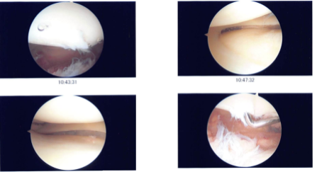
Arthroscopic images taken during the surgery

Arthroscopic images taken during the surgery

After one week the patient was seen in the office, she is here for her postoperative examination, no x-rays were needed. She denies fever, chills but her right shoulder is also hurting. She also developed rash over the neck, arm and chest wall since the last few days which has not been improving.

She took Benadryl also. Before discussing treatment, options and proceed with formal physical therapy as well as a home exercise program for rehabilitation of the shoulder.

We have decided to do the right RCT repair and that is needs to be planned for later, we also discussed the right knee arthroscopic chondroplasty, measurement of cartilage lesion and possible biopsy to be planned on next visit.

May need MACI in future, and the patient might see PCP for rash and restart Clindamycin for 5 days for discharge from surgical wounds. We went over the arthroscopic pictures and removed the stitches during the visits. We will continue with ice and elevation of the shoulder to decrease swelling and pain.

We will wean them off any narcotic medications and progress to anti-inflammatories and Tylenol as long as there are no contraindications to these medications. We also discussed the risk and benefits and common side effects of taking these medications. The doctor sees the patient back in three weeks’ time to evaluate her progress and discuss the next procedures.

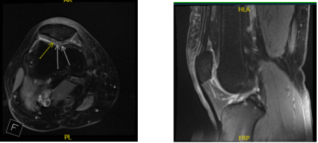
After one month the patient visits the office to do an MRI for another possible surgery, the MRI reviewed and discussed by the doctor, it showed that the patient had a subchondral fracture and cyst in the medial tibial plateau. We discussed treatment options and opted for surgical management.

We discussed risks and benefits including infection, bleeding, injury to adjacent nerves and vessels, need rehabilitation, need to be using crutches and non-weight bearing, and systemic complications including blood clot, cardiac, pulmonary, neurological complications including death. The patient understood and signed an informed consent.

MRI-3T Right shoulder non-contrast

MRI-3T Right shoulder non-contrast

MRI-3T Cervical Spine non-contrast

MRI-3T Cervical Spine non-contrast

The patient was taken to the operating room for a second time, where she was placed on a well-padded operating table. General anesthesia was induced. The left lower extremity was prepped and draped aseptically in the usual fashion. The left lower extremity was put on.

Tunica was applied on the right side and the leg was clamped. Draping was performed in aseptic fashion. Time-out was called. Tunica was elevated. Lateral entry portal was made with a parapatellar incision. Arthroscope was inserted and the median entry portal was made with the use of a spinal needle.

Examination of the medial femoral condyle showed lateral free-margin degenerative tear of the medial meniscus along with grade 4 osteoarthritic changes around medial tibial plateau extending from the anterior to the posterior surface along the region.

Examination of the medial intercondylar notch and lateral compartment showed no abnormality. Examination of the patellofemoral compartment showed 2 to grade 3 osteoarthritic changes of the lateral facet of the patella, which was debrided with the use of shaver. A biter was used for the meniscectomy on the medial meniscus. Microfracture chondroplasty was performed in multiple spots over the medial femoral condyle.

Final picture was taken and saved. Arthroscope was removed and plan for medial tibial plateau fixation was done. Incision was marked under fluoroscopy correlating with the MRI pictures.

Subchondroplasty needles x3 were inserted in the region and checked in the rotational fluoroscopy and drawn to be in acceptable position. A 3 cc of calcium triphosphate cement, which was premixed, was injected and trocars were inserted over the noodle to push that cement into the bone.

Pictures were taken to check and confirm the position of the cement. The cement was allowed to cure for about eight minutes after which the cannulas were removed and the area was cleaned. Picture was taken and saved again.

The incision was thoroughly irrigated and drained. Closure was done with the use of #2-0 subcu and # 3-0 nylon for the skin. A 20 cc of 0.5% Marcaine was injected into the knee. Dressing was done with the use of xeroform, 4 x 8, ABU, webril, and Ace wrap. The patient was extubated and moved to recovery in a stable condition.

The patient has been seen in the office multiple times in the past and has undergone arthroscopic surgery. She has been complaining of right shoulder pain for some time. We have tried cortisone injections in the AC joint as well as subacromial space, which have helped, but temporarily.

The patient has tried physical therapy also, it has helped temporarily. We discussed treatment options. We did an MRI, which showed a partial thickness rotator cuff tear along with acromial spurring, no meningeal arthritis.

The patient was seen by me multiple times in our office with complaints of injury, pain in both shoulders and both knees after a traffic accident. The patient had arthroscopic surgery on her knee and shoulder in the past.

At this time, she was complaining of right shoulder pain since her accident which has not improved despite cortisone injections and physical therapy. We did an MRI, which showed a partial rotator cuff tear. There was acromial spurring and AC arthritis.

Also, we discussed treatment options and opted for surgical management. We discussed risks and benefits and complications including infection, bleeding, injury to adjacent nerves and vessels, need for repeat surgery, need for rehabilitation, systemic complications including blood clot, cardiac, pulmonary, neurological complications among others. The patient understood and signed an informed consent.

The patient was taken to the operating room for the third time, where she was placed on a well-padded operating table. General anesthesia was induced. A supraclavicular nerve block was given in the holding area already. Preoperative antibiotic was given.

The patient was placed in a left lateral position with the right shoulder up. The right shoulder was prepped and draped aseptically in the usual fashion. Time-out was called. Bony prominences were marked over the skin and incision was given along the soft spot posteriorly to enter the glenohumeral joint.

A spinal needle was used to make an anterosuperior portal. Under arthroscopic guidance, an incision was given. The cannula was inserted along an anterosuperior portal.

A probe was inserted and examination of the glenohumeral joint was done, which showed extensive tearing of the labrum on the superior and the anterior part. There was tearing of the subscapularis tendon as well as intra articular tearing of the supraspinatus and the infraspinatus tendons.

A shaver was inserted from the anterosuperior portal and the debridement of the glenoid labrum, subscapularis, supraspinatus, and infraspinatus was performed. Pictures were taken and saved. There was no lesion on the glenoid cartilage.

The scope was inserted into the subacromial space and shaver was introduced and a subacromial bursectomy was done. There was acromial spurring. A Coblation wand was used followed by a burr to do an acromioplasty and remove the acromial spur along with release of the CA, coracoacromial ligament.

The 1-cm of distal clavicle was also excised with a burr. There was no rotator cuff tear seen on the bursal site. The shoulder was thoroughly irrigated and drained.

Final pictures were taken and saved. Closure was done with # 3-0 nylon. Dressing was done with Adaptic, 4×4, ABD, tape. The patient was put in a shoulder immobilizer and extubated and moved to recovery in a stable condition.

With regular visits and continued physical therapy and home remedies which are such a big help to the patient, she became well after all the surgery she went through.

Disclaimer – Patient’s name, age, sex, dates, events have been changed or modified to protect patient privacy.

I am Vedant Vaksha, Fellowship trained Spine, Sports and Arthroscopic Surgeon at Complete Orthopedics. I take care of patients with ailments of the neck, back, shoulder, knee, elbow and ankle. I personally approve this content and have written most of it myself.

Please take a look at my profile page and don't hesitate to come in and talk.