In shoulder arthroscopy, a tiny camera called an arthroscope is inserted into your shoulder joint by the surgeon. The word arthroscopy literally means “to look within the join”. Your surgeon uses the images from the camera to guide tiny surgical instruments as they are displayed on a video monitor.
Without surgery, rotator cuff injuries cannot heal, but many patients can benefit from nonsurgical therapy by gaining better functional mobility and reducing discomfort by strengthening their shoulder muscles. It’s not always necessary to have surgery just because there’s a rip.
A 46 year-old patient was in our office with complaints about pain in both shoulders, left and right and lower back following a car accident. He had no pain before the accident. He has tried PT and chiro with no relied. He is not able to perform activities of daily use.
The patient visited with an MRI result that showed at L3-4, there is disc bulge and left foraminal herniation. There is bilateral foraminal impingement more prominent on the left than on the right.
At L4-5, there is a disc bulge with bilateral foraminal impingement. At L5-S1, there is a broad-based central herniation with an annular tear directly abutting the bilateral S1 nerve roots.

MRI Lumbar Spine non-contrast
We discussed the treatment options for the patient’s diagnosis, which included living with the extremity as it is, organized exercises, medicines, injections and surgical options. We also discussed the nature and purpose of the treatment options along with the expected risks and benefits.
I educated the patient regarding the inherent and unavoidable risks which include, but are not limited to anesthesia, infection, damage to nerves and blood vessels, blood loss, blood clots, and even death were discussed at length.
We also talked about the possibility of not being able to return to prior activities or employment, the need for future surgery, and complex regional pain syndrome. The patient also understands there is a long rehabilitative process that typically follows the surgical procedure.
We talked about the possibility of not being able to alleviate all of the discomfort. Also, I explained there is no guarantee all the function and strength will return. The patient also understands the risks of re-tear or failure to heal.
The patient understands implants may be utilized during this surgery. The patient expressed understanding of these risks and has elected to proceed with surgery. We have discussed the surgical procedure as well as the realistic expectations regarding the risks, outcome, and post operative protocol.
The patient was taken to the operating room where he was placed on a well-padded operating table. Supraclavicular block was given by the anesthesia team. The patient was intubated. Preop antibiotic was given. The patient was turned in the right lateral position with the left shoulder up.
The patient was held in a bean bag in a sloppy lateral position. Left shoulder and arm were prepped and draped aseptically in usual fashion. Anterior portal was made posterior and medial to the posterior tip of acromion. Arthroscope was inserted from the posterior portal into the glenohumeral joint.
An anterosuperior entry portal was made with the use of spinal needle and cannula was Inserted. Examination of the joint showed fraying of the anterior margin of the glenoid as well as the glenoid labrum. There was minor fraying of the labrum at the attachment of the biceps also.
Examination of biceps showed no tenosynovitis or tears. Examination of the intra articular portion of the rotator cuff showed no tears. There was grade I, grade II osteochondral lesion of the posterior part of the head of humerus.
Shaver was inserted and debridement of the labrum as well as the glenoid and humeral head were performed. The arthroscope was entered from the anterior portal and the shaver entered from the posterior portal to complete the debridement.
Now, the arthroscope was entered into the subscapular space through the posterior portal. The shaver was entered from the anterosuperior portal and subacromial bursectomy was performed with the shaver.
Examination of the rotator cuff showed tear at the posterior margin of the supraspinatus with interstitial tearing intratendinous. Also shown was acromial spur and AC arthritis impinging on the rotator cuff. Acromioplasty was performed with the use of a thermal wand followed by 6.0 shaver.
Distal clavicular excision was also performed with the use of a thermal wand followed by burr. About 1 cm of distal clavicle was excised. Decision was made to repair the rotator cuff tear with the bioinductive implant from Smith & Nephew.
The bioinductive implant was inserted through the lateral portal into the joint. Superior portal was made for the insertion of tacks. The tacks were inserted through the superior portal and the bioinductive implant was tacked onto the rotator cuff with the use of PLA tacks x7. Finding it in satisfactory position and fixation, final pictures were taken and saved.
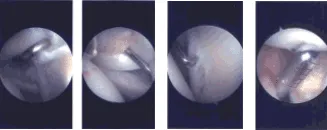
Intraoperative Arthroscopy Images
The shoulder was thoroughly irrigated and draped. Closure was done with use of 3-0 nylon. Dressing was done with the use of Xeroform, 4 x 8, ABD and dressing. The patient was put in a shoulder sling. The patient was recovered, extubated and moved to recover in a stable condition.
The patient was seen for post operative check up. We have decided to do formal physical therapy as well as a home exercise program for rehabilitation of the shoulder.
The patient did well after the surgery and continued physical therapy. Patient checked in for a follow up visit after a month and saw significant improvement on his shoulder.
Disclaimer – Patient’s name, age, sex, dates, events have been changed or modified to protect patient privacy.
The content on this page has been authored, edited, or approved by the doctors below, and was last reviewed for accuracy on October 21, 2025.
