An elbow bone spur is a bony protrusion that protrudes from the bone at the elbow joint. Some bone spurs are not apparent and may not cause symptoms; however, others may irritate your tendons, nerves, and muscles. This elbow inflammation may cause pain.
Osteophytes should be treated only if they cause pain or stiffness. Because osteophytes are closely related to arthritis, the treatments you may require are the same.
Patient is a 36 year-old male. Patient is here with complaints of right elbow pain. He states his elbow started to swell when he was playing basketball. He did not hit it or fall or injure it but it started to swell. He currently works as an NYPD.
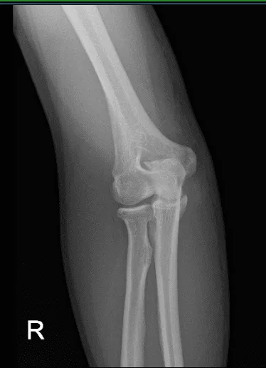
At first glance, it is suspected to have a radial head fracture. We did x-rays and found that he has a large osteophyte of the olecranon possibly secondary to chronic birth signs of the elbow.

X-ray of right elbow
We discussed treatment options and the patient opted for surgical management. We discussed the risks and benefits including infection, bleeding, recurrence of the osteophyte, injury to the adjacent nerves and vessels, elbow stiffness among others. The patient understood and signed an informed consent.
The patient was taken to the operating room and general anesthesia was induced. Preoperative antibiotics were given. Tourniquet was applied on the right upper extremity. Right upper extremity was prepped and draped aseptically in the usual fashion. Time-out was called.
A curvilinear incision was given over the point of elbow with the curve towards the radial site. With sharp dissection, the olecranon bursa was reached. The olecranon bursa was excised to its margins. The osteophyte could be reached.
Periosteum was elevated and Bovie was used to identify the osteophyte and delineated. An osteotome was used to excise the osteophyte. Once the osteophytes were excised, the margins were flushed thoroughly. No sharp edges were left.
The bursa was completely excised. Tourniquet was released and hemostasis was achieved. Closure was done in layers using #0 Vicryl, # 2-0 Vicryl and Monocryl. Dermabond was used followed by dressing in the form of 4 x 8s, Webril, Ace wrap. The patient was extubated and moved to the recovery unit in a stable condition.
He is here for his one week postoperative visit with X-rays right elbow. He Denies fever, chills and is able to return to work after a week of operation. X-ray showed that previous surgery is healing and soft tissue swelling overlying the olecranon process.
Postoperative X-ray of right elbow
We advised the patient to continue with ice and elevation of the knee to decrease swelling and pain. We also continue to utilize early mobilization and mechanical prophylaxis to reduce the chances of a deep vein thrombosis.
We will wean them off any narcotic medications and progress to anti-inflammatories and Tylenol as long as there are no contraindications to these medications. We also discussed the risk and benefits and common side effects of taking these medications.
And the patient will be back every three to four weeks to evaluate their progress. With continuous physical therapy and follow up, the patient gets well and able to be back on his normal routine.
Disclaimer – Patient’s name, age, sex, dates, events have been changed or modified to protect patient privacy.
The content on this page has been authored, edited, or approved by the doctors below, and was last reviewed for accuracy on October 19, 2025.
