Case Study: Shoulder Debridement and Mini-Open Biceps
Tenodesis performed to a 43 year-old male patient
Construction workers are physical workers too. Physical works that demand strenuous physical exertion for extended periods including walking, climbing, chopping, throwing, lifting, pulling and frequently carrying heavy objects weighing fifty pounds or more.
Patient presents with left shoulder pain that started due to overuse in construction work over time. He is not able to lift the arm and carry out ADL or do work with it. He feels very weak in the shoulder.
He saw a physician at Urgent care also. He has been trying home exercises but the weakness and pain has been progressing.
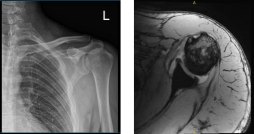
X-ray results showed no significant degenerative changes that’s why we decided to take an MRI. MRI impression as follows: Plate tear of the intra-articular portion of the long head of the biceps tendon and the large stump of proximal fibers displaced into the anterior medial joint recess.
The distal fragment is retracted into the upper arm distal to the pectoralis major attachment. There is moderate biceps tenosynovitis. Moderate subscapularis tendinosis with partial-thickness articular sided tearing at the insertion. Mild to moderate supraspinatus tendinosis.

MRI of the biceps
We discussed treatment options and the patient having failed conservative treatment, opted for surgical management.
We discussed risks and benefits including infection, bleeding, nonhealing, shoulder pain, need for rehabilitation, possibility of not finding tendon and not able to perform a tenodesis, systemic complications including blood clots, cardiac, neurological, and pulmonary complications including death.
The patient understood and signed the informed consent.
The patient was taken to the operating room where he was placed on a well-padded operating table. General anesthesia was induced. He was turned into the right lateral position with the left shoulder up.
Axillary roll was used under the axilla to protect the nerves. All the bony prominences were well padded. A bean bag was used to position him laterally. The left shoulder was prepped and draped aseptically in the usual fashion.
Preop antibiotic was given. The shoulder was put in 45-degree abduction and traction. Ten-pound weight was used. A time-out was called.
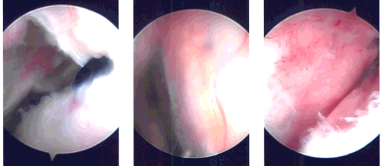
Entry portal was made posteriorly through the soft spot. An arthroscope was entered into the glenohumeral joint. There was considerable degeneration of the labrum as well as torn and frayed stumps of the biceps in the joint. An anterior entry portal was made using a Wissinger stick.
The cannula and shaver were introduced. The biceps tendon was shaved off to its origin. Labral tears were also shaved. There was inflammation around the subscapularis with synovitis which was also debrided. Examination of the rest of the joint showed intact cartilage of the glenohumeral joint.
There was partial bursal sided tearing of the rotator cuff in the region of the rotator interval which was debrided. The scope was now entered into the subacromial joint where there was minimal bursitis which was cleaned.
There was no spurring of the acromion. Decision was made not to do acromioplasty or distal clavicle excision. The scope was removed and biceps tenodesis was planned.
The left arm was removed from the traction and put into abduction and external rotation. A 4-cm incision was given along the lateral part of the inferior border of the pectoralis major. With sharp dissection, the deltoid fascia was cut.
Through the interval underneath the pectoralis major muscle, the soft tissues were dissected and the tendon of the long head of the biceps could be seen. It was delivered to the wound with the help of finger and right-angle forceps.
The tendon was retrieved with blunt force out of the wound. Once it was out, FiberLoop was used to whipstitch the tendon from the musculotendinous junction proximally. Excess of tendon was removed.
Now, the bicipital groove was reached under the pectoralis major. The retraction was done with Army-Navy and Hohmann retractors. The bone was cleaned with the use of Bovie and a bicortical drill was used to locate the site of insertion. Once it was in, the FiberLoop was tied over the button.
The unicortical drilling was done with a 7.5-mm drill bit. Once it was done, the drill was removed and the button was inserted over its positioner. Once the button was bicortically inserted through the humerus, it was ejected and locked.
Once it was locked, a limb of suture was passed through the tendon and tied over each other. Now, the limb of the suture was passed through the button and through the bioabsorbable screw and it was fixed unicortically over the tendon.
The suture was tied again. Final fixation was checked and found to be very sturdy. The wound was thoroughly washed and drained. Hemostasis was achieved.
Closure of the wound was done in layers using #0 Vicryl, # 2-0 Vicryl and Monocryl. Arthroscopic wounds were also sutured with Monocryl. Dressing was done using 4 x 8s, ABD and tape. The patient was put in a shoulder sling and moved to recovery in a stable condition after extubation.

Intraoperative Arthroscopy Images
Patient returned after a week post-operative. Post Operative Exam: General Appearance: no swelling or warmth; tenderness, passive motion limited, and active motion limited; and wound clean and dry and neurovascular intact. The patient is doing well after the surgery.
He has no other signs and symptoms and we decided to discussed the treatment options, we will proceed with the formal physical therapy as well as a home exercise program for rehabilitation of the shoulder.
We removed the stitches during the visit. We will continue with ice and elevation of the shoulder to decrease swelling and pain. We will continue to utilize early mobilization and mechanical prophylaxis to reduce the chances of a deep vein thrombosis.
We will wean them off any narcotic medications and progress to anti-inflammatories and Tylenol as long as there are no contraindications to these medications. We also discussed the risk and benefits and common side effects of taking this medication. the three weeks’ time to evaluate their progress.
Disclaimer – Patient’s name, age, sex, dates, events have been changed or modified to protect patient privacy

Dr. Vedant Vaksha
I am Vedant Vaksha, Fellowship trained Spine, Sports and Arthroscopic Surgeon at Complete Orthopedics. I take care of patients with ailments of the neck, back, shoulder, knee, elbow and ankle. I personally approve this content and have written most of it myself.
Please take a look at my profile page and don't hesitate to come in and talk.
