Case Study: Open Reduction and Internal Fixation
and Repair of the Lateral Ulnar Collateral Ligament
of the elbow in a 28 year-old patient
In order to replace the damaged ulnar collateral ligament within the elbow with a tendon from another part of the body, a procedure known as UCL reconstruction is frequently performed. Surgery’s objectives include stabilizing the elbow, minimizing or eliminating discomfort, and regaining stability and range of motion.
Additionally, excessive elbow use can result in tendinitis. Sprains, strains, fractures (broken bones), dislocations, bursitis, and arthritis are other causes of elbow discomfort. The reason will determine the course of treatment.
A 28-year-old patient presents left elbow pain. She was seen at UC where a long arm splint was applied. For aggravating factors, patients reported lifting and weight bearing.
For associated symptoms, swelling, tender to the touch, and pain with motion but reported no weakness, no numbness, no tingling, no redness, no warmth, no ecchymosis, no catching/locking, no popping/clicking, no buckling, no grinding, no instability, no radiation, no drainage, no fever, no chills, no weight loss, and no change in bowel/bladder habits.
For location, left. For quality, she reported aching and sharp. For severity, she reported severe.
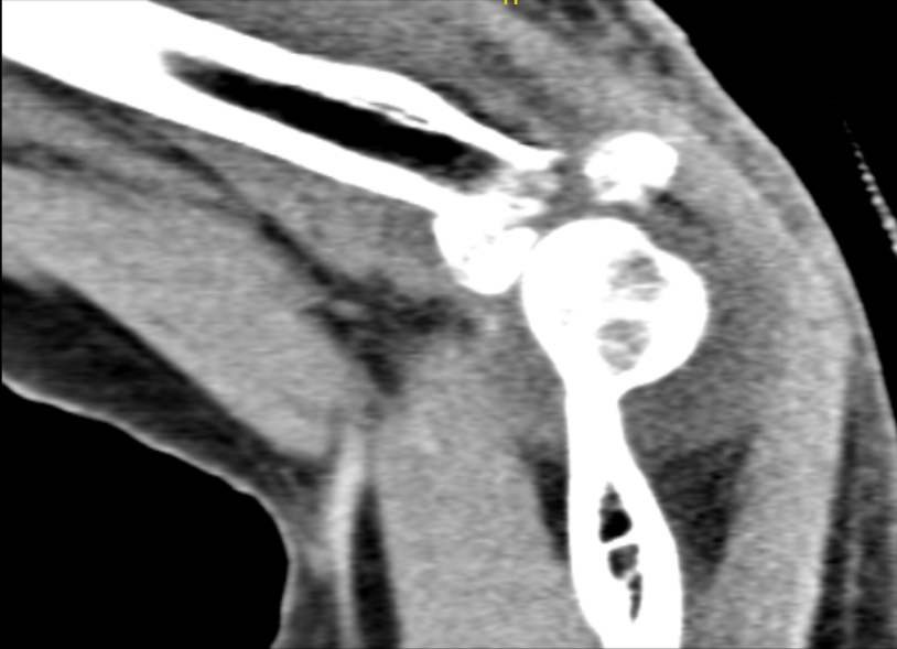
The patient presented a CT-scan result that showed Comminuted displaced acute fracture of the radial head as described above. Essentially nondisplaced acute fracture of the coronoid process of the ulna. Hemarthrosis.

CT Left Elbow Non-contrast
After the consent letter was verified the patient was taken to the operating room where anesthesia was induced. Antibiotics were given and a time out was given and the left upper extremity was prepped and draped in the sterile fashion. A Non Surgical tourniquet was placed. Esmarch was used to exsanguinate the limb to 250 mmHg.
A lateral incision was made of the elbow and dissection was carried down to the extensor apparatus. At this point the midportion of the lateral condyle was established as was the radial head, using fluoroscopy. An incision was made through the extensor sheath and the tendons were splint.
The supracondylar ridge was visualized and the muscles were elevated off the supracondylar ridge so that the joint could be visualized. The annular ligament was incised and the fracture hematoma was visualized and the radial shaft was visualized as well.
A partial tear of the ulnar collateral ligament was visualized as well as the fracture fragment. Once the fracture fragments were retrieved and the hematoma had been irrigated and suctioned, I began piecing together the fracture fragments in the back table using Kirschner wires to hold them in place.
Once these were in place I was then able to put the radial head onto the shaft of the radius and fired two Kirschner wires from the radial head to the shaft to hold this in place.
As soon as they were placed a Synthes radial head plate and shaft was placed secured with two Kirschner wires and placed proximally and distally. I was then able to place the remaining screws proximally and distally removing all the Kirschner wires at this point.
Final x-rays were obtained and good reduction was seen. The lateral collateral ligament was repaired using the bone tunnel and #2 FiberWire. The extensor apparatus was repaired with #2 FiberWire.
Vicryl was used as a running stitch on top. The deep dermal was then closed with 2-0 Vicryl and 3-0 Monocryl. Sterile dressings were placed. Splint was placed.
The patient was seen for post operative check up. We have decided to do formal physical therapy as well as a home exercise program for rehabilitation of the elbow. Patients regularly followed an office visit every 3-4 weeks.
Patient did well after the surgery and continued physical therapy. Patient checked in for a follow up visit after a month and saw significant improvement on her elbow.
Disclaimer – Patient’s name, age, sex, dates, events have been changed or modified to protect patient privacy.

Dr. Vedant Vaksha
I am Vedant Vaksha, Fellowship trained Spine, Sports and Arthroscopic Surgeon at Complete Orthopedics. I take care of patients with ailments of the neck, back, shoulder, knee, elbow and ankle. I personally approve this content and have written most of it myself.
Please take a look at my profile page and don't hesitate to come in and talk.
