The proximal and distal tibiofibular joints allow the fibula to move relative to the tibia, enhancing the ankle’s range of motion. The lateral malleolus also serves to support the ankle joint by forming the lateral wall of the talocrural joint.
The malleoli (“little hammers”) are the distal extremities of the fibula and tibia that overlap the talus. The distal end of the fibula is the lateral malleolus, while the medial and posterior malleoli are part of the tibia.
An interfragmentary screw, also known as a lag screw, is used to stabilize spiral or oblique fractures of a bone’s shaft.
A patient, a 24 year-old male, was seen in the office with pain in his ankle and foot because he fell on ice while returning from work. For aggravating factors, the patient reports standing, walking, lifting, twisting, weight bearing, exercise, and downstairs with associated symptoms of weakness, tingling, swelling, warmth, tender to the touch, and pain with motion. X-ray He was found to have a displaced rotated fracture of the lateral malleolus.
We discussed treatment options and the patient opted for surgical management. We discussed risks and benefits including infection, bleeding, implant failure, fracture failure, need for repeat surgery, injury to adjacent nerves and vessels, need for rehabilitation, blood clots, cardiac, pulmonary or neurological complications. The patient understood and signed an informed consent.
The patient was taken to the operating room where he was placed on a well-padded operating room table. General anesthesia was induced. Tourniquet was applied. Left lower extremity was prepped and draped aseptically in the usual fashion. Timeout was called.
An incision was begun over the lateral malleolus in a hockey stick fashion and was started in the line of incision. Fracture was exposed. Periosteum was elevated with the use of a periosteal elevator. The fracture was opened and irrigated. The fracture was reduced.
The fracture was found to be externally rotated and collapsed. The fracture was reduced and held with a clamp. Interfragmentary screw was applied from anterior superior to posterior inferior. A 3.5 anterior interfragmentary screw was inserted and the fracture was found to be safely reduced.
A lateral along the fracture and held with an Olive wire. X-ray showed satisfactory reduction in AP and lateral views. The plate was fixed to the distal fibula with the use of four distal locking screws and four proximally nonlocking screws.
Wound was thoroughly irrigated. Final pictures were taken and saved. Closure was done in layers using #0 Vicryl, #2-0 Vicryl, # 3-0 Monocryl. Dressing was done using 4×4, Webril, posterior splint and an Ace wrap. The patient was extubated and moved to recovery in a stable condition.
After a week from the surgery, the patient was sent for an X-ray. The AP, lateral and oblique. No prior examinations are available for comparison. Examination is performed through an overlying cast partially obscuring bony detail. The patient is status post ORIF distal fibular fracture.
The fracture line is barely visible. The surgical hardware is intact. Visualized aspects of the left talus, calcaneus and tarsal bones demonstrate intact cortical margins with no evidence of an acute fracture. There are no significant degenerative changes. Ankle mortise is well maintained.
There is no appreciable soft tissue swelling. There is no calcaneal plantar spur. It gives the impression that no prior examinations are available for comparison.
Examination is performed through an overlying cast partially obscuring bony detail. The patient is status post ORIF distal fibular fracture. The fracture line is barely visible. The surgical hardware is intact.
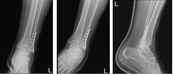
Left ankle X-ray complete 3
The patient was seen in the office for his post operative visits with an x-ray of his ankle that was reviewed by the doctor. He denies fever, chills, pain and NWB with crutches.
We agreed to go with conservative management for now, PT to be started, Ice and Elevation to decrease swelling and pain and OTC anti-inflammatory meds. We also advised to use Cam walker boots – can take off for showers, exercises only and NWB ambulation. For his Follow Up checkup, he will be back after 3 weeks.
After weeks the patient went back to the office for his follow up checkup, with an x-ray of his ankle for review. He denies fever, chills, pain and he is WBAT with boots, no crutches. X-rays were reviewed and discussed, there is a surgically treated fracture distal left fibula.
A faint fracture line is visualized, the visualized distal left tibia is unremarkable and visualized aspects of the left talus, calcaneus and tarsal bones demonstrate intact cortical margins with no evidence of an acute fracture.
Ankle mortise is well maintained however, there is mild soft tissue swelling laterally and no calcaneal plantar spur.
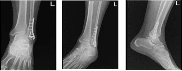
Left ankle X-ray complete 3
We agreed to continue with the medical treatment that we discussed before and the patient instructed to come back after 4 weeks for his follow up checkup.
With the continued follow up checkup the patient showed progress from time to time he visits the office. He gets well after the surgery and with the help of continuing physical therapy.
Disclaimer – Patient’s name, age, sex, dates, events have been changed or modified to protect patient privacy.
The content on this page has been authored, edited, or approved by the doctors below, and was last reviewed for accuracy on October 19, 2025.
