Case Study: Left Proximal Humerus Fixation
done to a 59 year-old male patient
Human arm bone is one of the longest bones we have on our body. It’s a very critical part of our ability to move our arms. It helps our muscles, tendons, ligaments and other parts of our circulatory system.
That’s the reason why we should take care of our humerus from any injury. One of the most common injuries that we may have is the proximal fracture that may be caused by a fall, car accidents and even sports.
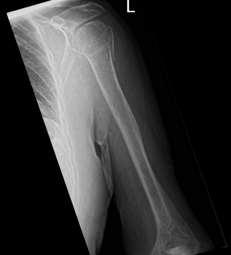
Patient seen today having complaints with his left shoulder. He stated that he fell off a ladder. And his left shoulder is the most affected one. He brings his X-ray with him. X-ray noted a comminuted fracture of the humeral head and neck and no change in position of fracture fragments compared to the left shoulder.

X-ray of the left shoulder
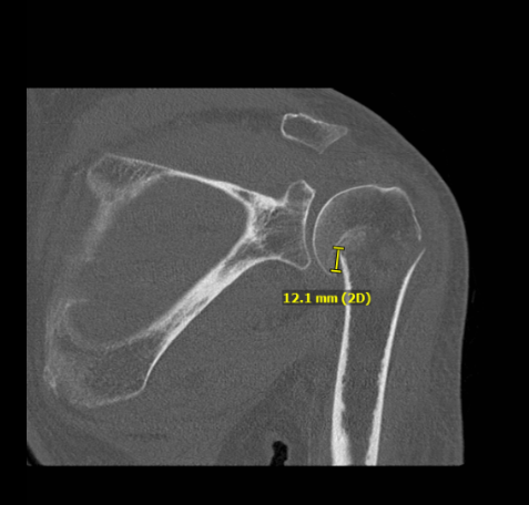
CT scan result also discussed to patient, comminuted proximal humeral fracture is impacted and slightly angulated fracture component at the surgical neck, slightly displaced fracture fragment at the greater tuberosity and a subtle nondisplaced fracture at the lesser tuberosity.
Bones are osteopenic. There is no evidence of bony bridging. There is a glenohumeral joint effusion.

We discussed the treatment options and the patient opted for surgical management. The patient has significant comorbidities and he had medical clearance along with cardiac and nephrological clearance.
We discussed the risks and benefits including infection, bleeding. nonhealing, deformity, stiffness, need for rehabilitation, need for shoulder replacement in future, need for repeat surgery among others.
We also discussed systemic complications including blood clot, cardiac, neurological, pulmonary complications including death especially considering his high comorbidities. The patient understood and signed informed consent.
The patient was taken to the operating room where he was placed on a well-padded operating table. General anesthesia was induced. The patient was put in a semi-reclining position. C-arm was brought in to see that the x-ray was visible in different views.
The patient was positioned appropriately and all bony prominences were well padded. Time-out was called. Preop antibiotic was given. The patient’s left upper extremity was prepped and draped aseptically in usual fashion.
Lateral incision was made from the mid acromion distally along the shaft of the humerus. With sharp and blunt dissection, the fascia was removed. The acromion tip was reached and a stay suture was put with 3-0 nylon at 4 cm from the acromial tip to restrict the dissection of the deltoid.
The deltoid fibers were split in line of the incision and the incision and the proximal humerus was reached. Subacromial bursa was reached and excised. The fracture was seen and cleaned. The fracture was reduced. A proximal humeral plate was chosen and inserted subperiosteally.
The axillary nerve could be palpated along the medial aspect of the deltoid. It was elevated and the plate was passed underneath it. Separate Incision was made for reaching the distal fragment of the plate. With sharp and blunt dissection, the plate was reached and held with the sleeves.
Finding the plate In acceptable position, a K-wire was used to hold the plate in position. Fixation of the distal fragment was done with the use of one nonlocking screw and followed by fixation of the proximal fragment in reduced position using multiple locking screws.
The fragment of greater trochanter was also reduced with #0 FiberWire and sutured to the plate. Distal excision was completed with the use of three cortical screws. Final pictures were taken and saved in multiple positions and found to be acceptable.
The wound was thoroughly cleaned and closed in layers using #0 Vicryl, # 2-0 Vicryl and Monocryl. Dressing was done using Dermabond, 4 x 4s, ABD, and Tegaderm. The patient was extubated and moved to recovery in a stable condition.
After one week, the patient returned for his post operative. Post Operative Exam: General Appearance: no swelling or warmth; tenderness, passive motion limited, and active motion limited; and wound clean and dry and neurovascular intact.
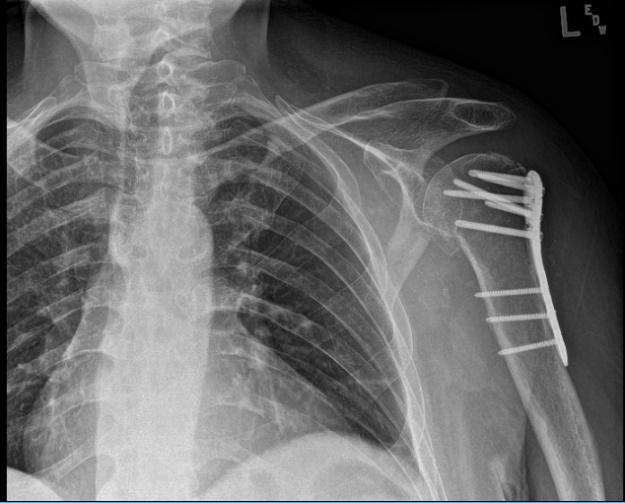
Xray reviewed- found surgically treated comminuted fracture of the humeral head and neck. By this time, we agreed to start Physical therapy.

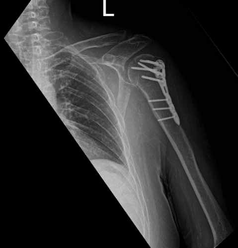
Post-operative X-ray.
We received a progress note from his physical therapist, the patient was seen 3 times in a week for 6 weeks. Per the initial evaluation, the patient is getting well from his surgery.
The patient is in status post-surgical repair of a comminuted fracture of the humeral head and neck. Fracture fragments are in good position and alignment. The surgical hardware is intact.

Patient last visit is 8 weeks after the operation, clearly seen on his X-ray that he is doing well with his surgery.
Disclaimer – Patient’s name, age, sex, dates, events have been changed or modified to protect patient privacy.

Dr. Vedant Vaksha
I am Vedant Vaksha, Fellowship trained Spine, Sports and Arthroscopic Surgeon at Complete Orthopedics. I take care of patients with ailments of the neck, back, shoulder, knee, elbow and ankle. I personally approve this content and have written most of it myself.
Please take a look at my profile page and don't hesitate to come in and talk.
