Surgery called chondroplasty is used to replace and remodel worn-out cartilage in joints. Degenerative cartilage is smoothed throughout the treatment, and any unstable cartilage flaps are trimmed. A delayed or non-healing union of a bone fracture can happen if it is not addressed.
The bone doesn’t mend at all in the first scenario; therefore, it will continue to be fractured. Because of this, swelling, soreness, and discomfort will keep getting worse.
A 34-year-old patient was in our office with complaints regarding left forearm and right knee pain due to a road traffic accident. He had a fracture of ulna, which had been treated nonoperatively.
He has been complaining of knee pain despite conservative management in the form of physical therapy and anti-inflammatory medications. The patient presented X Ray for his right knee that showed no significant degenerative changes. There are no fractures.
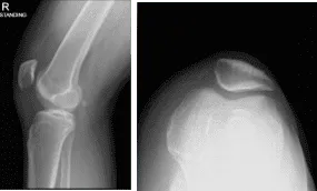
Right knee X-ray complete with Patella
We discussed the treatment options for the patient’s diagnosis, which included living with the extremity as it is, organized exercises, medicines, injections, and surgical options. I also discussed the nature and purpose of the treatment options along with the expected risks and benefits.
I educated the patient regarding the inherent and unavoidable risks which include, but are not limited to infection, stiffness, damage to nerves and blood vessels, blood loss possibly requiring transfusion, blood clots, persistent or worsening pain, loosening or failure of implants, instability, tingling or numbness, anesthesia and systemic complications including cardiac, pulmonary, neurological complications and even death were discussed at length.
I also talked about the possibility of not being able to return to prior activities or employment, the need for future surgery, and complex regional pain syndrome. The patient also understands there is a long rehabilitative process that typically follows the surgical procedure.
I talked about the possibility of not being able to alleviate all the discomfort. Also, I explained there is no guarantee all the function and strength will return. I discussed the type of implants that may be utilized during this surgery. The patient expressed understanding of these risks and has elected to proceed with surgery.
I discussed the patient’s medications and allergies and the possible need for medical and other clearances if needed. I have discussed the surgical procedure as well as the realistic expectations regarding the risks, outcome and post operative protocol.
The patient was brought to the operating room where general anesthesia was induced. The right knee was prepped and draped aseptically in the usual fashion after application of low thigh tourniquet and the foot in C- clamp. The tourniquet was elevated.
About 31 minutes of tourniquet time was used for the surgery. Preoperative antibiotics were already given before the inflation of the tourniquet. Lateral anterior portal incision was given through the parapatellar region. Arthroscope was inserted.
The medical entry was made with the use of a spinal needle and blade. Examination of the medial tibiofemoral compartment showed intact cartilage and meniscus. Examination of the intercondylar notch showed intact, but frayed and degenerated ACL. There was a tear flap from the anterior margin of the lateral meniscus.
A shaver was used to debride the meniscus and do the meniscectomy. Examination of the rest of the lateral tibiofemoral compartment showed intact cartilage of meniscus. Examination of the patellofemoral compartment showed grade 1 changes over the trochlea and patella.
There was a grade 2 osteochondral lesion of the patella, which was debrided with the use of a shaver. The rest of the knee was intact. The knee was thoroughly irrigated and drained. All the pictures were saved.
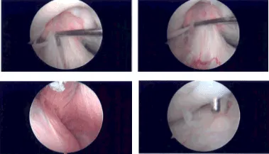
Intraoperative Arthroscopy Images
Closure was done with the use of 3-0 nylon. Then, 20 mL of 0.5% Marcaine mixed with 40 mg of Depo- Medrol was injected into the knee. The dressing was used with the use of Xeroform, Adaptic, 4×4, ABD, Webril and Ace wrap. The tourniquet was deflated.
The patient was sent to recovery in stable condition. The patient was seen for post operative check up. We have decided to do formal physical therapy as well as a home exercise program for rehabilitation of the knee. Patients regularly followed an office visit every 3-4 weeks.
Patient did well after the surgery and continued physical therapy. Patient checked in for a follow up visit after a month and saw significant improvement on his knee.
Disclaimer – Patient’s name, age, sex, dates, events have been changed or modified to protect patient privacy.
The content on this page has been authored, edited, or approved by the doctors below, and was last reviewed for accuracy on October 19, 2025.
