An open reduction and internal fixation (ORIF) are a surgical procedure that repositions shattered bone fragments. To hold the damaged bone together, screws, plates, sutures, or rods are employed. It’s a procedure used to support and mend a damaged bone.
A 59 year-old male patient visits the office complaining about his right ankle pain due to a fall for about two weeks. His right leg fell into a hole in the ground and got twisted. Upon the visits, we agreed to do a CT scan to find out what caused his right ankle’s pain.
CT-scan presented and reviewed; Bones are normally mineralized without a focal osseous lesion. There is a ununited obliquely oriented fracture of the distal fibula meta diaphysis extending to the level of the lower syndesmosis just proximal to the tibial plafond.
Distal fracture fragment measures 4 cm in length and is slightly posteriorly and laterally displaced less than 2 mm. There is no significant angulation. No other fractures are seen. There is a large os trigonum posterior to the talus with degenerative changes at the synchondrosis.
Ankle mortise is not widened. Talar dome is smooth without an osteochondral lesion. There is no tibiotalar joint space narrowing. Faint calcifications are seen along the lateral ligaments and within the lateral joint recess.
Mild chondrocalcinosis. There is no joint effusion. CT is suboptimal to evaluate the ligamentous complexes of the ankle.
There is mild narrowing of the posterior subtalar and talonavicular joints. Limited evaluation of the midfoot is unremarkable. Limited evaluation of flexor and extensor tendons are unremarkable. The Achilles tendon shadow is grossly unremarkable.
The unenhanced musculature is normal. Slightly displaced obliquely oriented fracture of the distal fibula extending to the lower syndesmosis just proximal to the tibial plafond Faint calcifications along the region of the lateral joint recess and lateral ligamentous favors mild chondrocalcinosis.

CT Right ankle non-contrast
Upon the result of the CT-scan, we started to discuss the treatment options including surgical and nonsurgical options. We also told the risks and benefits including infection, bleeding, implant failure or non healing, need for repeat surgery, injury to adjacent nerves and vessels, need for rehabilitation, ankle pain and instability.
However, the patient opted for surgical management. We also discussed risks, benefits, and complications including blood clots, cardiac, neurological, pulmonary complications including death. The patient understood and signed informed consent.
The patient was taken to the operating room where he was placed on a well-padded operating room table. Tourniquet was applied. The right lower extremity was prepped and draped aseptically in the usual fashion. Preoperative antibiotic was given. A time-out was called.
Right lateral incision was planned over the distal fibula. That incision was given. With sharp dissection, the bone and fracture were reached. The fracture was opened and washed. The fracture was reduced and held with clamps.
An interfragmentary 3.5 mm screw was used to compress the fragments. Once it was done, a mid-laterally driving tape was used to support the fixation. Distal locking as well as proximal nonlocking screws were used to achieve the fixation.
Intraoperative and final pictures were taken with the C-arm and saved. The wound was thoroughly washed. Closure was done in layers using #0 Vicryl, # 2-0 Vicryl and Monopril. Dressing was done. Tourniquet was removed. A short leg splint was placed. The patient was extubated and moved to recovery in stable condition.
A week after the surgery, the patient visits the office with an X-Ray result, and there is a surgically treated fracture of the distal fibula. Fixation plates and multiple screws are in place. Fracture fragments are in good position and alignment.
Right tibia demonstrates intact cortical margins with no evidence of an acute fracture. Visualized aspects of the right talus, calcaneus and tarsal bones demonstrate intact cortical margins with no evidence of an acute fracture. There are no significant degenerative changes.
Ankle mortise is well maintained and no appreciable soft tissue swelling. There is no calcaneal plantar spur. Status post ORIF distal fibular fracture. Fracture fragments are in good position and alignment. The patient is doing well after the surgery, elevating, and icing.
He still uses aspirin as pain relievers, he denies fever, chills but still he is NWB and using crutches. After two weeks, the patient’s skin peeling over the incision is getting better, the dressing performed with bacitracin, the splint is to continue for 2 weeks more. The patient was advised for 2 weeks for boot application.
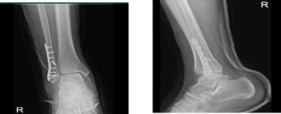
X-ray right ankle – 2 weeks after surgery
After three weeks from the surgery, the patient went to the office with X-ray results, for his follow up checkup. The skin of the patient peeling over the incision is resolved, the walking boot PWB 25%, to increase gradually with the help of the ice, elevation and OTC anti-inflammatory meds, the patient is doing very well after the surgery.
The surgical plate and screws transfix a fracture of the distal right fibula. Alignment is near anatomical, there is no significant interval change, the distal tibia is intact. There are no significant degenerative changes, the Ankle mortise is well maintained. There is no appreciable soft tissue swelling and no calcaneal plantar spur.
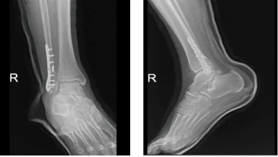
X-ray right ankle – 3 weeks after surgery
With the continued follow up checkup the patient showed progress from time to time he visits the office. He gets well after the surgery and with the help of continuing physical therapy.
Disclaimer – Patient’s name, age, sex, dates, events have been changed or modified to protect patient privacy.
The content on this page has been authored, edited, or approved by the doctors below, and was last reviewed for accuracy on October 18, 2025.
