Case Study: Medial Meniscectomy of Torn Meniscus
A torn meniscus cartilage in the knee can be treated with an outpatient, minimally invasive surgical technique called an arthroscopic meniscectomy. Injuries sustained when participating in sports frequently result in the meniscus being torn.
Meniscectomy is a frequent orthopedic operation used to treat meniscal disease in the elderly population that causes knee pain. Your tibia and femur may begin to inappropriately rub together without this meniscus cushion.
This may eventually result in ongoing knee pain. It might eventually lead to arthritis. As the cartilage “cap” deteriorates, the bones below begin to scrape against one another.
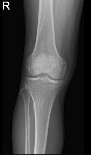
The patient is 66 years of age and came into the office with complaints of right knee pain. He remembered that he experienced the pain on and off a year ago. Patient denies remembering an injury that occurred. His knee X-ray showed no fractures and normal radiographs that’s why we agreed to take an MRI.

MRI of right knee
His MRI showed an impression as follows: Tricompartmental degenerative changes. Horizontal tear involving the body and posterior horn medial meniscus. Mild distal quadriceps tendinosis, without tears.
We discussed treatment options and the patient opted for surgical management. We discussed risks and benefits including infection, bleeding, nonhealing. need for repeat surgery, need for rehabilitation, development of arthritis and possible need for cortisone injections as well as total knee replacement in the future. The patient understood the risks and signed the informed consent.
The patient was taken to the operating room where he was placed on a well-padded operating room table. General anesthesia was induced. Preoperative antibiotic was given. Tourniquet was applied.
Right lower extremity was prepared and draped aseptically in the usual fashion. A time-out was called. A lateral entry portal was made and an arthroscope was entered.
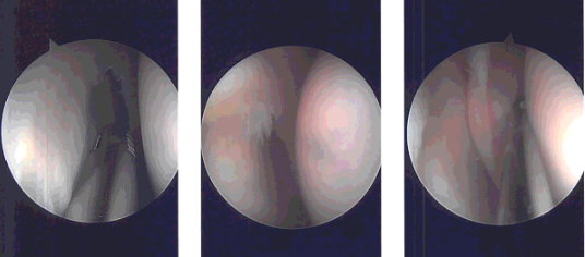
A medical entry portal was made using a spinal needle. Examination of the medial tibiofemoral compartment showed tears of the posterior body of the medial meniscus. The tear was cleaned using biters as well as shaver. were achieved. Examination of the Intercondylar notch showed intact ACL.
Examination of the lateral femoral condyle and lateral tibiofemoral compartment showed medial fraying of the lateral meniscus. Debridement was done using a shaver and biters.
Examination of the patellofemoral joint showed no chondral damage. There was chondral damage on the medial femoral condyle, which was cleaned using a shaver.
Final pictures were taken and saved. Closure was done using #3-0 nylon. Then, 9 cc of Naropin was mixed with Depo-Medrol 40 mg and injected into the knee. Dressing was done using 4 x 4, ABD, Ace wrap. The patient was extubated and moved to the recovery unit in stable condition.

Intraoperative Arthroscopy Images
He returned to our office a week after his operation. No X-ray required for his visit. He is doing well. He is not using crutches and not limping. Denies pain, fever, chills. Activities as tolerated and can now return to his work. Patients get well with continuous follow up and with the help of physical therapy.
Disclaimer – Patient’s name, age, sex, dates, events have been changed or modified to protect patient privacy.

Dr. Vedant Vaksha
I am Vedant Vaksha, Fellowship trained Spine, Sports and Arthroscopic Surgeon at Complete Orthopedics. I take care of patients with ailments of the neck, back, shoulder, knee, elbow and ankle. I personally approve this content and have written most of it myself.
Please take a look at my profile page and don't hesitate to come in and talk.
