Shoulder arthroscopy is a minimally invasive surgical technique used to treat various shoulder problems. It enables the surgeon to visualize, diagnose, and treat shoulder conditions with small incisions, making it less invasive than traditional open surgery. This procedure is widely used to address a variety of shoulder issues, including sports injuries, arthritis, and tendon problems.

X-ray showing the normal shoulder anatomy.
How Common It Is and Who Gets It? (Epidemiology)
Shoulder arthroscopy is a common procedure for patients suffering from shoulder pain that doesn’t respond to conservative treatments like physical therapy or medications. It is particularly effective for conditions like rotator cuff tears, shoulder instability, arthritis, and impingement syndromes. Athletes, active individuals, and older adults are more likely to require this procedure due to overuse injuries, age-related wear and tear, or traumatic injuries.
Why It Happens – Causes (Etiology and Pathophysiology)
The shoulder is a highly mobile joint that is prone to injury. Overuse, trauma, and degenerative diseases like arthritis can lead to damage of the rotator cuff tendons, labrum, and cartilage. Conditions like frozen shoulder or tendonitis may also require arthroscopic intervention. The procedure helps to treat these issues by allowing the surgeon to remove damaged tissue, repair torn tendons, and restore normal function.
How the Body Part Normally Works? (Relevant Anatomy)
The shoulder joint consists of the humerus (upper arm bone), scapula (shoulder blade), and clavicle (collarbone). The ball of the humerus fits into the shallow socket of the scapula, forming a ball-and-socket joint. This configuration provides a large range of motion. Ligaments, tendons (like the rotator cuff), and the labrum stabilize and support the joint, allowing for smooth movement and flexibility.
What You Might Feel – Symptoms (Clinical Presentation)
Common symptoms of shoulder issues that may require arthroscopy include:
- Pain: Especially when moving the arm or lifting objects.
- Stiffness: Difficulty in moving the shoulder through its full range of motion.
- Weakness: Reduced strength or instability when lifting or rotating the arm.
- Swelling: Inflammation around the shoulder joint, often accompanied by tenderness.
How Doctors Find the Problem? (Diagnosis and Imaging)
The diagnosis of shoulder conditions is made through a combination of:
- Physical Examination: Assessing range of motion, strength, and tenderness.
- Imaging: X-rays are used to identify structural issues like fractures or arthritis, while MRIs or CT scans help visualize soft tissue damage, such as rotator cuff tears or labral injuries.
- Arthroscopy: In some cases, the procedure is performed for both diagnosis and treatment.
Classification
Shoulder arthroscopy is used to treat a variety of conditions classified as either:
- Rotator Cuff Tears: Damage to the tendons that stabilize the shoulder.
- Labral Tears: Tears in the cartilage that cushions the shoulder socket.
- Frozen Shoulder: A condition where the shoulder becomes stiff and painful.
- Arthritis: Degenerative changes in the shoulder joint leading to pain and loss of motion.
Other Problems That Can Feel Similar (Differential Diagnosis)
Conditions such as shoulder fractures, dislocations, or other soft tissue injuries can present with similar symptoms to those requiring arthroscopy. Tendonitis and bursitis are also common causes of shoulder pain that may need to be differentiated from more serious conditions that require surgical intervention.
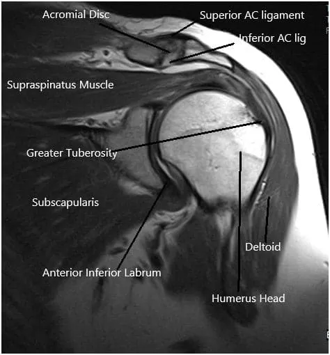
MRI of the shoulder joint.
Treatment Options
Non-Surgical Care:
-
- Physical Therapy: Exercises to strengthen the shoulder and improve mobility.
- Medications: Pain relievers, anti-inflammatory drugs, or corticosteroid injections.
- Rest and Ice: Used to reduce swelling and promote healing.
Surgical Care:
-
- Shoulder Arthroscopy: Minimally invasive surgery to repair or remove damaged tissues, such as the rotator cuff or labrum, and address impingement or arthritis.
Recovery and What to Expect After Treatment
Following shoulder arthroscopy, patients typically recover faster than with open surgery. Recovery involves:
- Post-operative Care: The shoulder is usually immobilized in a sling for a few days to weeks, depending on the type of repair.
- Rehabilitation: Physical therapy is essential to regain strength and mobility. Full recovery may take several months, depending on the complexity of the surgery.

Intraoperative arthroscopic images.
Possible Risks or Side Effects (Complications)
- General Risks: Infection, bleeding, and nerve injury are potential complications with any surgery.
- Specific Risks: Implant failure, stiffness, or further damage to the shoulder joint. In very rare cases, complications like damage to the blood vessels or nerves surrounding the joint may occur.
Long-Term Outlook (Prognosis)
Most patients experience significant improvement in pain and shoulder function after arthroscopic surgery. The procedure generally results in quicker recovery and fewer complications compared to open surgery. However, some patients may experience a recurrence of symptoms, especially if rehabilitation is not properly followed.
Insurance & Cost
Arthroscopy – Shoulder Joint at Complete Orthopedics is covered by Medicare and most major insurance carriers (Aetna, Anthem BCBS, Cigna, Empire BCBS, UnitedHealthcare), as well as most workers’ compensation and no-fault insurance plans. Your out-of-pocket cost depends on your specific plan, deductible, and the medical necessity criteria that apply to your case.
Call our billing team at (631) 981-2663 before scheduling to verify your coverage and discuss expected out-of-pocket costs. For the full list of carriers we accept and patient billing protections, see our Insurance Information page.
Frequently Asked Questions (FAQ)
Q. What is Shoulder Arthroscopy?
A. Shoulder arthroscopy is a minimally invasive surgery where a small camera and surgical instruments are used to treat conditions inside the shoulder joint.
Q. What conditions are treated with Shoulder Arthroscopy?
A. Conditions like rotator cuff tears, shoulder instability, labral tears, arthritis, and frozen shoulder are commonly treated with arthroscopy.
Q. How is Shoulder Arthroscopy performed?
A. A small incision is made in the shoulder, and a camera is inserted to visualize the joint. Instruments are used to treat the condition, such as repairing or removing damaged tissue.
Q. What are the benefits of Shoulder Arthroscopy?
A. The procedure is minimally invasive, with smaller incisions, less pain, quicker recovery, and fewer complications compared to open surgery.
Q. What are the risks of Shoulder Arthroscopy?
A. Risks include infection, bleeding, nerve damage, and stiffness. However, these complications are rare.
Q. What is the recovery time after Shoulder Arthroscopy?
A. Recovery typically takes several weeks to a few months, depending on the severity of the condition and the type of surgery performed.
Q. Can Shoulder Arthroscopy be performed on both shoulders at the same time?
A. It is usually performed on one shoulder at a time to ensure proper healing and minimize complications.
Q. Is physical therapy required after Shoulder Arthroscopy?
A. Yes, physical therapy is essential to restore strength, flexibility, and range of motion after surgery.
Q. Will I experience any loss of motion after Shoulder Arthroscopy?
A. Some temporary stiffness may occur, but with proper rehabilitation, most patients regain full or nearly full motion.
Q. How long does Shoulder Arthroscopy surgery take?
A. Most arthroscopic shoulder surgeries take 1-2 hours, depending on the complexity of the procedure.
Q. Can I walk immediately after Shoulder Arthroscopy?
A. Yes, since the procedure is minimally invasive, walking is usually possible immediately, though heavy lifting or overhead activities should be avoided.
Q. How effective is Shoulder Arthroscopy for treating shoulder problems?
A. Shoulder arthroscopy is highly effective in treating various shoulder conditions, with most patients experiencing significant pain relief and improved function.
Q. Are there any long-term complications of Shoulder Arthroscopy?
A. Long-term complications are rare but can include further damage to the shoulder joint, re-injury, or stiffness.
Q. How soon can I return to sports after Shoulder Arthroscopy?
A. High-impact activities should be avoided for several months, but light activities can often be resumed after a few weeks.
Q. Can Shoulder Arthroscopy be combined with other shoulder surgeries?
A. Yes, shoulder arthroscopy can be combined with other procedures like tendon repair or ligament reconstruction depending on the patient’s needs.
Summary and Takeaway
Shoulder arthroscopy is a minimally invasive procedure that can effectively treat a variety of shoulder conditions, providing faster recovery, less pain, and fewer complications compared to open surgery. Whether it’s for rotator cuff tears, labral injuries, or arthritis, this procedure allows for significant improvement in shoulder function and quality of life. Patients typically recover quickly, especially with proper rehabilitation, and can return to daily activities and sports faster than with traditional surgery.
Clinical Insight & Recent Findings
A recent study reviewed techniques to improve visualization during shoulder arthroscopy, a common minimally invasive procedure. The research highlighted various factors impacting surgical visibility, including patient positioning (beach chair vs. lateral decubitus), blood pressure control, and fluid management with arthroscopic pumps.
Notably, the use of epinephrine (EPI) and tranexamic acid (TXA) in the irrigation fluid was found to significantly enhance visual clarity. EPI acts as a vasoconstrictor, reducing bleeding, while TXA prevents fibrinolysis, contributing to better visualization and reduced post-operative complications.
The study concluded that these measures, along with careful positioning, play critical roles in optimizing the surgical environment, potentially improving outcomes and reducing complications. (“Study on shoulder arthroscopy – see PubMed.”)
Who Performs This Treatment? (Specialists and Team Involved)
Orthopedic surgeons specializing in shoulder and upper extremity surgery perform shoulder arthroscopy. In some cases, a multidisciplinary team, including physical therapists and rehabilitation specialists, may be involved in postoperative care.
When to See a Specialist?
If you experience persistent shoulder pain, stiffness, or instability that doesn’t improve with conservative treatments, it’s important to consult an orthopedic surgeon to evaluate whether shoulder arthroscopy is a suitable treatment option.
When to Go to the Emergency Room?
You should seek emergency care if you experience severe shoulder pain, swelling, or a sudden loss of function, especially after a trauma or injury.
What Recovery Really Looks Like?
Recovery from shoulder arthroscopy involves a few weeks of rest, followed by physical therapy to restore strength and mobility. Most patients experience a return to normal activities within a few months, depending on the type of surgery performed.
What Happens If You Ignore It?
Ignoring shoulder problems can lead to worsening pain, decreased function, and further damage to the shoulder joint. Early intervention with procedures like arthroscopy can prevent these issues and improve long-term outcomes.
How to Prevent It?
Maintaining shoulder strength through regular exercise, proper lifting techniques, and avoiding overuse can help prevent shoulder injuries and conditions that may require surgery.
Nutrition and Bone or Joint Health
A diet rich in calcium and vitamin D supports bone and joint health, which is important for preventing shoulder issues and aiding recovery after surgery.
Activity and Lifestyle Modifications
After surgery, low-impact activities are ideal for maintaining fitness while the shoulder heals. Avoid overhead activities and heavy lifting until cleared by your surgeon.
The content on this page has been authored, edited, or approved by the doctors below, and was last reviewed for accuracy on July 2, 2026.
