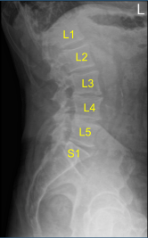
Case Study: Durotomy Repair
The patient is in status post L2-L3 right sided hemilaminectomy and microdiscectomy for a right L3 nerve root compression done two weeks back. The patient was well postoperatively. Their pain was resolved. They were able to do the usual activities of daily living. They were having some constipation. They also did some yard work.
One week after the surgery, They started having clear watery drainage from the wound. Couple of days later, They started having headaches. They were seen by me in the office yesterday where we suspect it to be a CSF leak. We already started oral antibiotics. We discussed the need for an MRI and followed by possible exploration and closure of the CSF leak.
The patient understood. We discussed options of treatment as well as risks and benefits including infection, bleeding, injury to adjacent nerves and vessels, CSF leak, recurrent bleed, need for repeat surgery, and need for rehabilitation amongst others. The patient was then signed an informed consent.
The patient was met by us in the holding area and taken to the operating room. General anesthesia was induced. They were placed in a prone position on the Jackson table with Wilson frame. The back was prepped aseptically using Betadine. Draping was performed. Time-out was called.
Preop antibiotic was given. The wound was opened. All the sutures were removed. The wound was debrided. Operating microscope was brought in. We reached the laminotomy where some loose tissue was there. There were some spicules which were not readily sharp.
Further decompression of the laminotomy was performed with exposure. Foraminotomy of the L3 nerve root was also performed. The CSF leak was found on the dorsal surface. It was about 5 mm long. It was packed using patties and then Surgicel.
Revision decompression was completed by removing the superior L3 Lamina, medial, lateral and inferior L2 lamina using kerrison punch No 3. The inferior facet of the L2 vertebrae on the right side was loose and had to be removed. After thorough decompression, the incidental durotomy was planned for repair.
The microscope was used for the decompression as well as the durotomy repair. A #6-0 Prolene was used and two sutures were put around the ends. Once the closure was performed satisfactorily, a Valsalva was given twice, raising the pressure to 40 mmHg. No leakage was found.
The repair was covered with patties and low pressure pulse lavage was performed using bibiotic and normal saline. The patties were removed. The repair was sealed with layers of Surgicel and Evicel. Hemostasis was achieved. Once decompression and hemostasis was deemed satisfactory, closure was done in layers.

Post-Op
The patient followed up 2 weeks after the surgery. They were able to return to all their regular daily activities. Their initial complaint of Right L3 radiculopathy had been solved. There was no more leakage of Cerebrospinal fluid.
Disclaimer – Patient’s name, age, sex, dates, events have been changed or modified to protect patient privacy.

Dr. Nakul Karkare
I am fellowship trained in joint replacement surgery, metabolic bone disorders, sports medicine and trauma. I specialize in total hip and knee replacements, and I have personally written most of the content on this page.
You can see my full CV at my profile page.
