Case Study: Management of Avascular Necrosis of the Left Hip with Total Hip Replacement in a 25-year-old Male with Prior Core Decompression Surgery
A young 25-year-old male presented to our office with complaints of worsening left groin pain. The patient reported being seen by another orthopedic surgeon and was diagnosed with avascular necrosis of the left head of the femur. He had previously undergone core decompression surgery of the left hip performed by his previous surgeon.
As per the patient, the pain in the left hip started insidiously about two years ago with no precipitating injury or fall. The patients reported taking corticosteroid medications (prednisone) for an extended period of time (6 months) before the onset of pain. The corticosteroid medications were prescribed for nephrotic syndrome.
The patients described a dull aching constant pain in the left groin region. The pain is worse with activities such as walking, navigating stairs, turning, standing, sitting, bending, squatting, etc. The pain decreased in intensity on rest but is not completely relieved.
The patients reported no radiation of the pain or any associated swelling, fever, redness, or rise in temperature of the left groin. The patient works as a construction worker and faces significant difficulty in his day-to-day activities.
He also noticed increasing difficulty in performing activities such as getting up from the toilet seat and climbing stairs. The patient reported he is unable to take nonsteroidal anti-inflammatory medications due to his kidney condition. He reports no relief with acetaminophen and is averse to the temporary use of any kind of opioid medications.
The medical history of the patients is significant for nephrotic syndrome and high blood pressure. The patient’s current medications include amlodipine, prednisone, metoprolol, and benazepril. He has a prior surgical history of core decompression of the left hip performed a year ago. He is a non-smoker and denied any long-term alcohol intake.
On physical examination, the gait of the patient was bipedal, unassisted, steady, and coordinated. There were exaggerated lumbar lordosis and functional/structural scoliosis. The bilateral shoulders, anterior superior iliac spines, patella, and medial malleolus were at the same level.
The skin overlying the left hip was normal with no signs of redness, sinus tracts, scar, discharge, swelling, inguinal lymphadenopathy, or local rise in temperature. There were small scars present on the outer side of the upper left thigh consistent with the percutaneous core decompression.
Mild tenderness was present in the left anterior hip joint line on deep palpation. The greater trochanter was not enlarged and non-tender. There was no evidence of any limb length discrepancy. The range of motion of the left hip was complete in all ranges with mild pain on abduction and internal rotation.
The examination of the spine, right hip, bilateral knees, and ankles was normal. There was no evidence of any neurological deficit in the bilateral lower extremities. The bulk, tone, and power of the lower extremities were unremarkable.
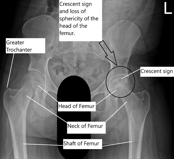
On radiological investigations, the X-ray of the left hip revealed avascular necrosis of the head of the femur with subchondral collapse visible in the form of a crescent sign. The X-ray of the right hip revealed a normal radiograph.

X-ray of the pelvis with both hip joints showing AVN of the left head of the femur.
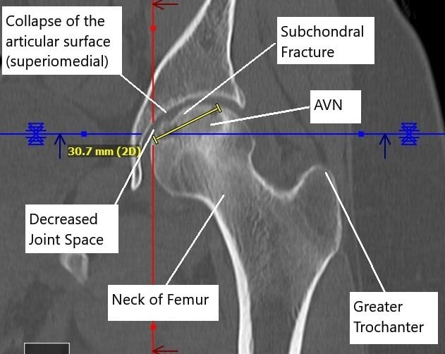
A CT without contrast revealed progression of the avascular necrosis since the core decompression surgery. Avascular necrosis involved the majority of the femoral head, most pronounced superomedially and measuring 3.1 x 3.8 cm.
There was a subchondral fracture superomedially and a mild collapse of the articular surface. There was a mild narrowing of femoral acetabular joint space superomedially with a suggestion of a small joint effusion.

Non-contrast CT image showing AVN changes in the left head of the femur.
The MRI of the left hip joint revealed a status-post core-decompression for left femoral head AVN. There was marrow edema surrounding the necrotic bone and suggestion of a subchondral “crescent sign” along with the superomedial femoral head, indicating fracture and stage III AVN. There was a slight focal collapse of the femoral head in this location.
The patient was advised a total hip replacement in the view of Ficat & Arlet stage 3 osteonecrosis/avascular necrosis of the head of the femur with collapse. The risk, benefits, and potential complications were explained to the patient’s satisfaction. The patient understood the possibility of a revision surgery given his young age.
A total hip replacement of the left hip was performed using an acetabular shell of 56 mm with a 36 mm polyethylene liner. A ceramic head was used with a size nine extended offset tapered wedge-designed uncemented stem.


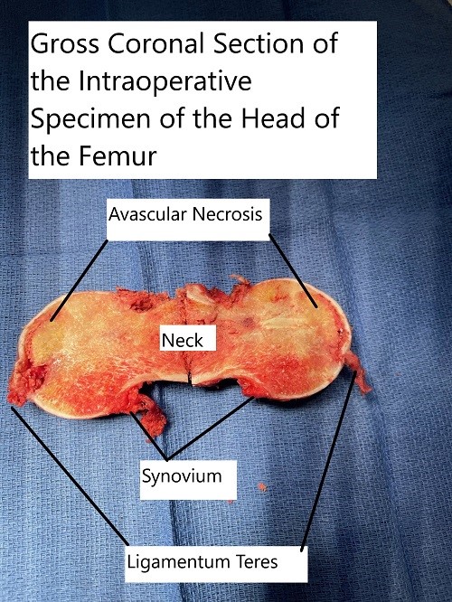
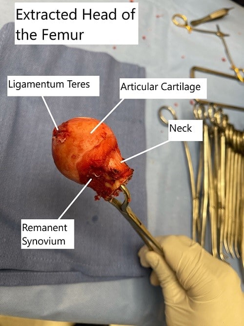
Intraoperative extracted head of the femur with a coronal cut section showing the avascular necrotic area in the superior medial head.
The patient had an excellent recovery post-op, and there was minimal pain. The patient was given aspirin for deep vein thrombosis prophylaxis. The patient was advised to bear weight as tolerated. The sutures were removed 14th post-op day after the wound was inspected to be clean, dry, and intact.
The patient was able to participate in physical therapy and returned to his baseline activities quickly. He was advised regular follow-up for signs of avascular necrosis in the right hip joint. The patient was relieved to be free of pain and to resume the activities he enjoyed.
Disclaimer – Patient’s name, age, sex, dates, events have been changed or modified to protect patient privacy.

Dr. Suhirad Khokhar
My name is Dr. Suhirad Khokhar, and am an orthopaedic surgeon. I completed my MBBS (Bachelor of Medicine & Bachelor of Surgery) at Govt. Medical College, Patiala, India.
I specialize in musculoskeletal disorders and their management, and have personally approved of and written this content.
My profile page has all of my educational information, work experience, and all the pages on this site that I've contributed to.
