A 74 year old male presents with chronic right hip pain which began in approximately May of 2017. Patient complains that the pain is affecting is gait and weight bearing, and hindering his quality of life.
The pain severity makes him unable to walk, stand, or sit for any extended period of time. The patient has underwent extended periods of physical therapy, injections and massage therapy with no substantial or lengthy relief.

Patient is in overall good health besides a heart murmur in the past. Pre- Operative films show severe arthritis on the right side. I discussed surgical options with the patient, the patient’s best option was to undergo a right hip total arthroplasty; Patient was overall excited to undergo the procedure, so he could get back to a pain-free life. The patient was retired and although he did not have to transport to and from employment, he did have 4 grandchildren that he actively cared for, He was very motivated to be able to move around with them.
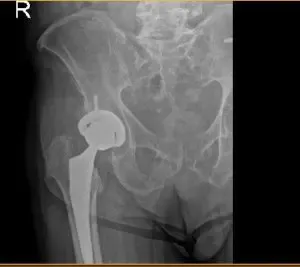
Post op hip X-ray
The patient underwent a right total hip arthroplasty, A curved incision centered over the greater trochanter was used for the arthrotomy. Skin and subcutaneous tissues were incised. Fascia was then divided. The hip was placed into internal rotation and the posterior soft tissue structures were then taken down and tagged for future repair.
The hip was then dislocated. The lesser trochanter to the center measurement was taken and the neck resection was made at the correct level. Head was then removed. Attention was then turned towards the acetabulum.
The remainder of the labrum was then debrided. The acetabulum was sequentially reamed. The final shell was then placed into position in the correct abduction and anteversion. Screw was used for additional fixation. Additional osteophytes were then removed.
The poly was placed over the shell. Attention was turned towards the femur and the femur was sequentially broached. The final broach was left into position. The head was then placed over the trunnion. Hip was then relocated and trialed through a full physiological range of motion. The hip was stable in all physiological range of motion. Hip was then dislocated.
The combined anteversion was correct. The hip was then dislocated. The trial components were removed. Final components were then placed into position.
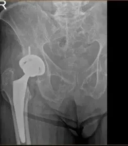
X-ray of a hip replacement
The hip was then relocated and the posterior soft tissue structures were then taken down and tied for future repair. A thorough lavage was given. Injection was given. Fascia was then closed with Eithibond.
Cutaneous tissues were closed with Vicryl. Subcuticular tissues were closed with 2-0 Vicryl. Skin was closed using Monoderm. Sterile dressing was then applied over the wound and the patient was then transferred to the postoperative care unit in stable condition.
Post-operative films show Right hip total arthroplasty, in good alignment, mild sacroiliac degenerative changes. Patient returned for a post operative appointment for staple removal and patients prognosis is good, due to an uncomplicated surgery. Patient is compliant with the outpatient physical therapy program, and is weight bearing fully bilaterally.
Patients physical therapy services were concluded with excellent results. Patients stated his quality of life was significantly improved, and can now move far better than he could even after his original procedure. Patients home activities are completed with ease. The patient is enjoying his life and is now able to do activities with his grandchildren.