Synovial Facet Cyst
A synovial facet cyst is a fluid-filled sac originating in the facet joint of the spine. The fluid-filled sac is most commonly found in the lower spine (lumbar spine). Although most synovial facet cysts may not cause any symptoms, they may compress the nerve roots and cause radiating leg pain. The condition is a result of degenerative changes in the lower spine.
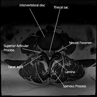
The facet joints are present above and below each vertebra. They are formed where the superior and the inferior articulating facets of the adjoining vertebrae meet. A synovial joint is a joint consisting of a tight fluid-filled capsule. The synovial fluid is generated by the capsule’s inner lining, which lubricates and nourishes the facet joint.

MRI axial section showing the facet joint.
With growing age, there may be degenerative changes in the spinal column. The water content of the intervertebral disc may decrease which may lead to decreased disk height. The loss of height of the disk leads to degeneration and instability of the facet joint, known as facet joint syndrome. The bones forming the joint may rub against each other, causing pain in the lower back.
The synovial fluid formation is increased by the body to help lubricate the facet joint. At times, the extra fluid may get caught in the lining of the capsule and form a fluid-filled sac. The fluid-filled sac may encroach upon the precarious space in the neural foramen and cause pressure symptoms such as spinal canal stenosis.
The fluid buildup in the cyst occurs slowly but may happen fast if there is bleeding inside the cyst (facet cyst hematoma). The majority of the facet cyst formation occurs in the L4-L5 spinal level.
The patients may complain of low back pain. The back pain may be associated with radiating leg pain. At times, patients may complain of feeling numbness and tingling sensation in the legs. The symptoms may progress suddenly in the case of synovial facet hematoma.
Neurogenic claudication may occur in patients with synovial facet cyst. The patients may complain of pain in the buttocks and the legs, which is worse on walking. The pain is relieved at rest by bending forward. The space in the neural foramen is reduced on bending backward and standing. The effect is exacerbated with a synovial facet cyst encroaching the space.
The diagnosis is made on an MRI with clinical examination eliciting nerve root defects. The physician may perform a thorough neurological test to look for the nerves’ motor and sensory function. The synovial facet cyst is visible on MRI as an MRI is able to differentiate between various soft tissue structures.
The management of synovial facet cyst is mainly conservative. The conservative management consists of nonsteroidal anti-inflammatory medications to relieve pain and inflammation. Physical therapy is initiated to improve muscle strength and flexibility. Heat and cold therapy may help relieve the pain and stiffness in some patients.
Epidural injections may also help in the temporary control of pain. In patients where the symptoms clearly correlate with the synovial facet cyst, a CT guided cyst rupture and cyst steroid injection may be done. Repeat injections may be required in some patients.
Operative management is indicated in patients with persistent symptoms despite conservative management. Surgical management may be done in the form of lumbar laminectomy and decompression. The surgery consists of creating an opening in the lamina and the facet joint to take out the cyst. Laminectomy and decompression surgery are generally done in patients with unilateral symptoms.
Facetectomy with fusion surgery is the treatment of choice when indicated. The surgery is indicated in patients with bilateral symptoms, recurrence following laminectomy and decompression, and central canal stenosis.
Do you have more questions?
What exactly is a synovial facet cyst?
A synovial facet cyst is a fluid-filled sac that develops in the facet joints of the spine due to degeneration, often causing compression of nearby nerves.
What is the difference between a synovial cyst and a ganglion cyst?
Synovial cysts are lined by a synovial membrane and contain joint fluid, whereas ganglion cysts lack this membrane and are filled with gelatinous material. Both can cause similar symptoms.
How do synovial cysts form?
These cysts form when the synovial fluid, which lubricates the joints, escapes into small tears in the joint capsule, often due to degenerative changes like osteoarthritis.
Are lumbar synovial facet cysts common?
No, they are relatively rare but are more frequently diagnosed due to advancements in imaging technologies like MRI. Posterior cysts are more common than anterior ones.
Are all lumbar synovial facet cysts symptomatic?
No, many cysts are asymptomatic and may only be discovered incidentally during imaging for other conditions.
What are the primary symptoms of a lumbar facet cyst?
Symptoms can include lower back pain, leg pain (radiculopathy), numbness, tingling, muscle weakness, and in severe cases, difficulty walking due to nerve compression.
Can a lumbar synovial cyst resolve on its own?
In some cases, small cysts may spontaneously resolve, but this is not common. Larger cysts usually require medical intervention.
What causes lumbar synovial facet cysts to grow?
They grow due to continuous degenerative changes in the spine, particularly in the facet joints, and the ongoing production of synovial fluid in response to joint irritation.
Is there a risk factor for developing these cysts?
Age, osteoarthritis, spondylolisthesis, and previous spinal trauma are significant risk factors, as these conditions contribute to joint degeneration.
How is a lumbar synovial cyst diagnosed?
The gold standard for diagnosis is MRI, which provides detailed images of the soft tissues, including cysts, and helps differentiate them from other spinal abnormalities like herniated discs.
Can lumbar synovial cysts mimic other conditions?
Yes, they can mimic conditions like disc herniations, spinal stenosis, or other nerve compressions due to similar symptoms like radiculopathy and lower back pain.
What conservative treatments are available for lumbar synovial cysts?
Conservative treatments include physical therapy, NSAIDs, corticosteroid injections, and using a back brace to reduce symptoms without surgery.
What is percutaneous aspiration or rupture of the cyst?
This is a minimally invasive procedure where a needle is guided into the cyst to either aspirate the fluid or rupture the cyst under imaging guidance, followed by a steroid injection.
Is percutaneous treatment effective?
It can be effective in the short term, but studies show that about 29% of patients need repeat procedures or further surgery because the cyst recurs or symptoms return.
When is surgery recommended for a synovial facet cyst?
Surgery is usually recommended when conservative treatments fail or if the cyst is causing significant nerve compression, leading to pain, weakness, or neurological deficits.
What is the difference between decompression surgery and decompression with fusion?
Decompression surgery removes the cyst or bone tissue compressing the nerves, while decompression with fusion involves stabilizing the spine by fusing two vertebrae together to prevent further instability.
What are the surgical options for treating lumbar synovial cysts?
Surgical options include decompression surgery, where part of the bone or tissue compressing the nerve is removed, and sometimes fusion surgery is added if there is spinal instability.
What are the risks of surgery for a lumbar synovial cyst?
Risks include infection, bleeding, spinal instability (especially if fusion is not done), and potential injury to nearby nerves or the spinal cord, though these complications are rare.
What is the recovery time after surgery for a lumbar synovial cyst?
Recovery time varies depending on the type of surgery. Decompression surgery usually involves a shorter recovery (4-6 weeks), while decompression with fusion may take several months for full recovery.
Can the cyst come back after surgery?
Recurrence is uncommon after surgical removal, particularly if fusion is also performed. However, in rare cases, cysts can form at other levels of the spine.
Is there any way to prevent synovial facet cysts from forming?
While there’s no guaranteed way to prevent these cysts, maintaining spinal health through regular exercise, a healthy weight, and avoiding heavy lifting can reduce the likelihood of degenerative changes in the spine.
What is the prognosis for patients with lumbar synovial facet cysts?
The prognosis is generally good, especially for those who undergo surgery. Most patients experience significant relief from pain and neurological symptoms.
Does the size of the cyst determine the treatment approach?
Not necessarily. Treatment is more often based on symptoms rather than cyst size. Even a small cyst can cause significant symptoms if it compresses a nerve.
What type of doctor treats lumbar synovial facet cysts?
Orthopedic surgeons or neurosurgeons specializing in spine surgery typically treat synovial facet cysts, especially when surgery is necessary. Other specialists, such as pain management doctors, may provide non-surgical treatments.
Will I need physical therapy after surgery for a synovial cyst?
Yes, physical therapy is often recommended after surgery to help restore mobility, strengthen the muscles supporting the spine, and reduce the risk of future spinal issues.

Dr. Suhirad Khokhar
My name is Dr. Suhirad Khokhar, and am an orthopaedic surgeon. I completed my MBBS (Bachelor of Medicine & Bachelor of Surgery) at Govt. Medical College, Patiala, India.
I specialize in musculoskeletal disorders and their management, and have personally approved of and written this content.
My profile page has all of my educational information, work experience, and all the pages on this site that I've contributed to.
