Cauda Equina Syndrome
At Complete Orthopedics, we specialize in addressing back pain through personalized treatment plans and a range of surgical options. We emphasize thoroughly understanding your symptoms to provide accurate diagnoses and effective treatments to alleviate pain following surgery or to assess the necessity for further surgical procedures.
Our clinics are located throughout New York City and Long Island, connected to six top hospitals, and equipped with the latest technology for advanced back surgeries and outstanding orthopedic care. Scheduling a consultation with our orthopedic experts is simple, either online or by phone.
Overview
Cauda equina syndrome (CES) is a surgical emergency caused by compression of the lower spine’s thecal sac. The condition may lead to weakness and numbness of the legs, bowel bladder dysfunction, and the saddle area’s numbness.
The management is urgent surgical decompression and which is preferably performed within 48 hours since the onset of symptoms.
Anatomy of the Spine
The spinal cord transmits the signal to and from the body to the brain as it travels down the vertebral column. Enclosed in the vertebral canal, the spinal cord is protected by the vertebral column.
The spinal cord gives numerous branches known as nerve roots that unite to form spinal nerves.

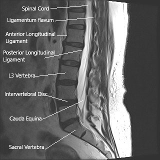
MRI of the lumbar spine in sagittal section showing cauda equina (horse’s tail)
The spinal cord ends at the lower level of T12 or L1 vertebrae. At its termination, the spinal cord forms a collection of spinal nerves L1-S5 that continues down the vertebral canal and exiting at various neural foramina.
This collection of spinal nerves is known as Cauda Equina (horse’s tail).
The nerves in Cauda Equina control the movement of the lower limbs and maintain the bladder bowel control. The nerves also transmit the sensory signals from the lower extremities and the saddle area to the brain.
The nerves in Cauda Equina are more sensitive to any compression compared to other nerves in the body.
Cauda Equina Syndrome Causes & Pathology
Intervertebral disc herniation is the most common cause of cauda equina syndrome. The herniated disc may compress the thecal sac leading to compression of the spinal nerves. Spinal canal stenosis may also lead to compression of the cauda equina.
Trauma due to falling from a height or motor vehicle accident may cause dislocation, collapse, or retropulsion of fracture fragments in the canal. The subsequent compression leads to cauda equina syndrome. Misplaced vertebrae, known as spondylolisthesis, may lead to narrowing of the vertebral canal.
Other causes of cauda equina syndrome include spine tumors, epidural hematoma (blood collection in the outer layer of the dural sac), or inadvertent injury during spinal surgery.
The mechanical pressure on the nerve roots leads to the decreased blood supply to the nerves. The resulting ischemia leads to damage to the nerve fibers, leading to loss of the neural signal transmission.
Cauda Equina Syndrome Symptoms
The most common initial complaint is back pain, usually followed by leg pain in one or both legs. The patients may complain of numbness in the perianal/saddle area. The patients often complain of not being able to feel the toilet paper. Patients may also complain of sexual dysfunction.
There may be a weakness in one or both of the legs. The patients may also complain of numbness and tingling sensation in the legs. In advanced cases, there may be loss of bladder control.
The patients may feel a sense of incomplete evacuation or involuntary evacuation of the bladder. In rare cases, the patients may experience loss of bowel control as well.
Cauda Equina Syndrome Diagnosis
The physician makes the diagnosis of cauda equina syndrome after a detailed history and physical examination. The physician may perform a thorough neurological analysis to ascertain the motor and sensory functions of the limbs and the peri-anal area.
An MRI is the imaging study of choice for evaluating neurological compression. An X-ray is usually done in traumatic injury cases and to assess the bony structures along with MRI. A CT myelography may be done in patients who are unable to do an MRI evaluation.
Blood investigations may be done if an infection is the suspected cause of cauda equina syndrome. Bladder investigations such as urodynamic studies may be done to assess the filling volumes of the bladder before and after voiding.
Cauda Equina Syndrome Treatment
Cauda Equina syndrome is a surgical emergency, and the standard management is decompression surgery. The decompression surgery may involve discectomy, discectomy with laminectomy, or discectomy with laminectomy and fusion.
The surgical technique involved in the management depends on the patient’s anatomy and the compressing structures.
In discectomy surgery, the herniated intervertebral disc is removed to alleviate the pressure on the spinal nerves. The discectomy is usually performed after creating a hole in the lamina (laminotomy).
In cases of spinal stenosis, discectomy is often done along with lumbar laminectomy surgery. The surgeon may also add implants along with lumbar fusion surgery if instability is suspected.
Cauda Equina Syndrome Complications
Complications occur in untreated cauda equina syndrome cases or in cases where the surgery was delayed for more than 48 hours. The symptoms of cauda equina syndrome may persist or may recover very slowly over the years.
Bladder dysfunction may require a permanent catheterization of the bladder. Sexual dysfunction, chronic pain, and weakness of the lower extremities may persist for years.
As with any surgery, there may be potential complications associated with decompression surgery, such as nerve root injury, bleeding, infection, blood clots, dural tear, epidural fibrosis, etc.
Preventing Cauda Equina Syndrome
While not all cases of CES can be prevented, there are steps you can take to reduce your risk:
Maintain a Healthy Weight: Excess weight can put additional strain on your spine.
Exercise Regularly: Regular physical activity helps strengthen the muscles that support your spine.
Practice Good Posture: Proper posture can reduce the risk of spinal problems.
Lift Safely: Use proper lifting techniques to avoid injuring your back.
Avoid Smoking: Smoking can contribute to degenerative changes in the spine.
Conclusion
Cauda equina syndrome is a serious condition that requires immediate medical attention. Understanding the symptoms, causes, and treatment options can help ensure that you receive prompt and effective care if you or someone you know develops CES. With timely treatment, many people can recover and regain function, but early diagnosis and intervention are crucial.
If you experience symptoms of CES, such as severe lower back pain, leg weakness, or loss of bladder or bowel control, seek medical help immediately. Early treatment can make a significant difference in outcomes, helping to prevent permanent damage and improve the quality of life.
Do you have more questions?
What is Cauda Equina Syndrome?
Cauda equina (Latin) means horse tail. It is a name given to the nerve roots in the lumbosacral spinal canal as they look similar to horse tail on visualization. Cauda equina syndrome is the compression of the spinal nerve roots in the lumbar and sacral area of the spine Lesions above this level leads to compression of spinal cord and is not cauda equina syndrome, but the presentation is more dramatic and carries same or more urgency as of cauda equina syndrome.
Compression of the spine causes weakness of upper or lower extremities with increased reflexes and with or without involvement of the bowel or bladder. Cauda equina syndrome is essentially a clinical presentation of new onset or worsening weakness in one or both lower extremities, gait abnormality, involvement of the bladder and numbness in either lower extremity and peri genital area (sacral anesthesia).
These patient may also have sexual dysfunction. The patients usually have severe back pain. Cauda equina syndrome is usually associated with pain in the back and occasionally with radiculopathy. Rarely, patients with cauda equina syndrome may present without any complaints of pain.
This happens due to compression of the nerve roots in the lumbar spine and leading to dysfunction of the muscles as well as altered sensation that are taken care by the specific nerve roots. This is a severe form of presentation of nerve root compression in the lumbar spine.
It can present acutely or over many months or days. It may be caused due to degeneration of disk fragment, mass in the spinal canal, bleeding in the spinal canal, intraspinal mass like tumor, fracture, gunshot or rarely with a birth defect (usually an arteriovenous malformation). The presentation can be acute or chronic depending on the pathology.
What injuries can cause cauda equina syndrome?
Fractures or dislocations of the lumbosacral spine may lead to cauda equina syndrome. These are traumatic injuries and are associated with high velocity accidents like motor vehicle accident or fall from height. Traumatic disc herniation may also lead to cauda equina syndrome.
What type of physicians take care of cauda equina syndrome?
Acute cauda equina syndrome is usually treated under the care of a spine surgeon who can be of orthopedic or a neurosurgical background. A chronic cauda equina syndrome in which a surgery has been ruled out is usually under the care of neurologist and may also need care of oncologist or radiation oncologist in cases which are associated with malignancy or metastasis.
Why is rectal exam needed in cauda equina syndrome?
The rectal exam can be of diagnostic value in cauda equina syndrome lacks rectal sphincter is associated with cauda equina syndrome and should be checked in all patients. It may be the only sign of Cauda Equina Syndrome.
How to diagnose a cauda equina syndrome?
Cauda equina syndrome is diagnosed clinically due to its characteristic presentation of new onset or worsening of weakness, gait abnormality, bowel or bladder dysfunction, sexual dysfunction and sacral anesthesia. Confirmation of diagnosis is done with advanced imaging specifically. MRI which helps to find out the level of compression as well as helps in diagnosing the pathology.
In patients who have contraindications for MRI (Pacemaker, aneurysmal clip), CT scan and myelogram may be done. Confirmatory diagnosis of the pathology can only be done at the time of surgery and with the need of histopathologic examination of the tissue compressing on the nerve roots.
What are the causes of cauda equina syndrome?
Causes of acute cauda equina syndrome can be a disk fragment (most common), fracture or dislocation of the spine, a hematoma caused by bleeding in the spinal canal, vascular insult to the nerve root due to underlying systemic or local pathology, infection, inflammation, gunshot or stabbing to spine, motor vehicle accident or fall, birth defect (arteriovenous malformation).
Cause of chronic cauda equina syndrome can be a slow growing mass or a degenerative spine with disk fragment or hypertrophied ligaments causing lumbar stenosis, birth defects etc. A mass can be in the form of tumor or metastasis or rarely a primary tumor of the nerve roots or the nerve elements.
Can I be disabled due to cauda equina syndrome?
Cauda equina syndrome is a disabling disease. It leads to weakness and usually with dysfunction of the bladder and sometimes bowels too. It leads to impaired gait due to the weakness of the muscles of the leg. Due to involvement of bladder, it may lead to retention or incontinence of urine leading to use of alternate methods for evacuation of the bladder. Patients may have gait problems too.
How do I know I have a cauda equina syndrome?
Patients with cauda equina syndrome usually have new onset or worsening weakness in one or both lower extremities, gait abnormality, involvement of the bladder and numbness in either lower extremity and peri genital area (sacral anesthesia). These patient may also have sexual dysfunction. The patients usually have severe back pain. These patients may have preexisting back pain and radiculopathy. Patients may have a history of cancer with or without metastasis to the spine and may have already undergone treatment for that in the past.
What do I do if I have cauda equina syndrome?
An acute onset cauda equina syndrome is a surgical emergency and the patient should go to the ER immediately. Advanced imaging should be performed as soon as possible to confirm the diagnosis after the physical examination of the patient. If a cauda equina syndrome is confirmed, a surgery may be needed to decompress the spine and allow the recovery of the nerve roots. Patients with chronic cauda equina syndrome who have insidious onset over many days or weeks, should seek medical attention to confirm the diagnosis as well as plan a possible treatment for their disease.
How common or rare is cauda equina syndrome?
Cauda equina syndrome is a rare presentation of various pathologies of the spine. Most pathologies present with back pain or/and radiculopathy. They may also develop subtle weakness, but developing profound weakness with involvement of bladder and gait is rare. It is even rarer in degenerative disk disease and lumbar canal stenosis.
What is the treatment of cauda equina syndrome?
Patients with acute presentation of cauda equina syndrome with confirmatory diagnosis on an MRI showing mass effect on the nerve roots usually will need an urgent or emergent surgery to decompress the nerve roots. They will need to be admitted to the hospital and will need to undergo physical rehabilitation for optimization of the function as well as enhance their recovery.
Patients with chronic cauda equina syndrome may also need surgery depending on the pathology, but may also need adjuvant treatment especially in the cases of malignancy or metastasis in the form of chemo or radiotherapy. Occasionally these patients with chronic cauda equina syndrome can manage with adjuvant treatments only without the need for surgery. Patients with poor general condition and multiple comorbidities may have to be treated non-surgically so as to curtail the risk to their life due to the anesthesia as well as the surgery.
How is the recovery from cauda equina syndrome?
Recovery from cauda equina syndrome depends on the type of pathology, amount of compression, number of levels involved as well as the surgery performed. In most cases, the recovery will happen if their condition has been treated promptly but may not lead to full recovery of the functions. Patients will need to undergo physical rehabilitation to optimize their function as well as enhance their recovery.
Can cauda equina syndrome cause bladder problems?
Cauda equina syndrome usually causes bladder problems in the form of retention or incontinence. These patients need to be treated for their bladder problems separately so as to allow recovery and at the same time avoid complications due to the condition.
Can cauda equina syndrome cause constipation?
Cauda equina syndrome can occasionally cause involvement of bowels also which may lead to constipation in most cases.
Can cauda equina syndrome cause death?
Cauda equina syndrome causes disability in the form of weakness of the lower extremities and involvement of bowel or bladder, and problems with ambulation, but it cannot be a direct cause of death, though in patients with chronic sequelae of cauda equina complications like deep vein thrombosis causing pulmonary embolism, urinary tract infection causing sepsis pulmonary infection or respiratory failure may be secondary cause of death in such patients.
Can you get cauda equina syndrome twice?
Cauda equina syndrome in itself is a rare entity and to get it twice is rarer, though not impossible. Patients who are predisposed to cauda equina syndrome like those with malignancy or metastasis or those with blood disorder and are on anticoagulants may rarely have cauda equina syndrome twice too.
Is cauda equina syndrome permanent?
An acute presentation of cauda equina syndrome if treated appropriately can lead to good recovery, but if not treated appropriately or in patients with chronic cauda equina, the sequelae of cauda equina syndrome may be long lasting or permanent too.
Can you get cauda equina syndrome with fusion surgery?
Any surgery on lumbar spine carries a risk of cauda equina syndrome. This can happen due to any bleeding at the surgical site, which leads to hematoma formation and compression of the nerve roots causing the presentation of cauda equina syndrome. These patients need to be treated urgently with decompression and need to be carefully followed up.
How to avoid or prevent cauda equina syndrome?
As the cauda equina syndrome and itself is a rare entity, there is no possible way to prevent a cauda equina syndrome. Patients who are on anticoagulants carry a higher risk of cauda equina syndrome, but the benefits of anticoagulant therapy far outweighs the risk of cauda equina syndrome or any other such bleeding complication. Similarly patient with metastases are at increased risk of cauda equina syndrome and their tumor is appropriately treated with chemo or radiotherapy, but prophylactic treatment with the surgery or radio or chemotherapy just to prevent cauda equina syndrome is not advisable.

Dr. Suhirad Khokhar
My name is Dr. Suhirad Khokhar, and am an orthopaedic surgeon. I completed my MBBS (Bachelor of Medicine & Bachelor of Surgery) at Govt. Medical College, Patiala, India.
I specialize in musculoskeletal disorders and their management, and have personally approved of and written this content.
My profile page has all of my educational information, work experience, and all the pages on this site that I've contributed to.
