Open discectomy (OD) and microdiscectomy (MD) are both surgical techniques used to treat lumbar radiculopathy (sciatica) caused by a herniated or bulging intervertebral disc. While both procedures aim to relieve nerve compression by removing disc material, they differ in their approach, surgical time, recovery time, and risks. The decision to choose between the two is influenced by the patient’s condition, the surgeon’s experience, and other factors such as the level of herniation.
How Common It Is and Who Gets It? (Epidemiology)
Lumbar disc herniation is a common cause of sciatica, with a high prevalence in adults between 30 and 50 years old. The condition is particularly common in individuals who engage in activities that strain the lower back, such as heavy lifting or prolonged sitting. Approximately 10-15% of people with lumbar disc herniation will require surgery, with microdiscectomy and open discectomy being the two most common surgical interventions.
Why It Happens – Causes (Etiology and Pathophysiology)
A herniated disc occurs when the soft, inner portion (nucleus pulposus) of an intervertebral disc pushes through a tear in the tough outer layer (annulus fibrosus). This herniation can compress spinal nerves, leading to symptoms like radiating leg pain (sciatica), numbness, tingling, or weakness in the lower extremities. Over time, the spine undergoes degenerative changes, which can contribute to disc bulging and nerve compression. Repetitive stress, trauma, or poor posture can also contribute to the development of disc herniation.
How the Body Part Normally Works? (Relevant Anatomy)
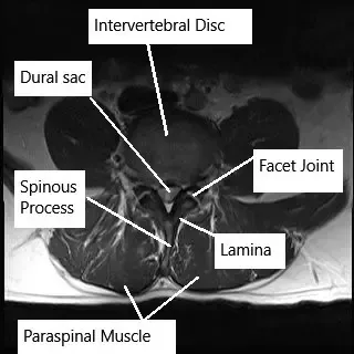
The lumbar spine consists of five vertebrae (L1-L5) separated by intervertebral discs. These discs act as shock absorbers and allow for flexibility and movement of the spine. The spinal cord runs through the spinal canal, and nerves branch out through openings between the vertebrae. These spinal nerves control the sensation and movement of the lower body, including the legs, feet, and pelvic organs. When a disc herniates, it can compress these nerves, leading to symptoms such as pain, numbness, and weakness.
What You Might Feel – Symptoms (Clinical Presentation)
- Radicular Pain (Sciatica): Pain that radiates from the lower back down the buttocks and into the legs.
- Numbness or Tingling: Sensation of “pins and needles” in the legs or feet.
- Weakness: Difficulty lifting the legs or feet, or feeling of instability.
- Back Pain: Localized pain in the lower back, often worsened by activity or movement.
- Loss of Reflexes: Reduced reflexes in the legs due to nerve compression.
How Doctors Find the Problem? (Diagnosis and Imaging)
- Physical Examination: To evaluate reflexes, strength, and areas of pain or numbness.
- MRI (Magnetic Resonance Imaging): The most effective tool for visualizing herniated discs and nerve compression.
- CT Scan: Provides a more detailed image of the bones and joints.
- X-rays: To rule out other causes of back pain such as fractures.
- Electromyography (EMG): To assess nerve function and confirm compression.
Classification
Open discectomy and microdiscectomy are both used to treat herniated discs, but they differ in terms of the approach:
- Open Discectomy (OD): Involves a larger incision and more extensive tissue manipulation to remove the herniated disc.
- Microdiscectomy (MD): A minimally invasive procedure using a small incision, often with an operating microscope or endoscope, to remove the herniated disc with less tissue disruption.
Other Problems That Can Feel Similar (Differential Diagnosis)
Conditions that may mimic the symptoms of lumbar disc herniation include:
- Spinal Stenosis: Narrowing of the spinal canal, often leading to similar symptoms of leg pain and numbness.
- Piriformis Syndrome: Compression of the sciatic nerve by the piriformis muscle in the buttocks.
- Sacroiliac Joint Dysfunction: Pain in the lower back and buttocks that may radiate to the legs.
- Facet Joint Syndrome: Degeneration of the facet joints, causing pain similar to sciatica.
Treatment Options
Non-Surgical Care
- Physical Therapy: Strengthening the back and abdominal muscles to reduce pressure on the spine.
- Medications: NSAIDs or corticosteroids for pain and inflammation relief.
- Epidural Steroid Injections: To reduce inflammation around the nerve roots.
- Nerve Blocks: To target and alleviate pain in specific areas.
Surgical Procedures
Open Discectomy (OD)
- Procedure: Involves a midline skin incision, retraction of paravertebral tissues, partial laminectomy, and removal of the ligamentum flavum to expose the spinal cord and nerve roots. The disc material is then removed, and the posterior longitudinal ligament is opened.
- Duration: Significantly shorter operation time (37.82±7.15 minutes).
Microdiscectomy (MD)
- Procedure: Utilizes a microscope for better visualization, with minimal laminectomy and removal of the ligamentum flavum. The nerve root is retracted medially, and the disc contents are removed.
- Duration: Longer operation time (49.07±6.88 minutes).
Recovery and What to Expect After Treatment
- Open Discectomy: Recovery time is generally longer due to the larger incision and more extensive tissue manipulation. Patients may need to stay in the hospital for 1-2 days and can expect a return to normal activities in 4-6 weeks.
- Microdiscectomy: Recovery is faster due to the minimally invasive nature of the procedure. Most patients can return to light activities within 1-2 weeks, with full recovery typically taking 4-6 weeks. Patients can usually go home the same day of the surgery.
Possible Risks or Side Effects (Complications)
- Open Discectomy:
- Infection: At the surgical site.
- Nerve Injury: Rare, but can occur during the surgery.
- Recurrent Herniation: The disc may herniate again at the same level.
- Dural Tear: Accidental tear of the dura mater, requiring repair.
- Microdiscectomy:
- Infection: Low risk, but possible.
- Nerve Injury: Less common due to better visualization.
- Recurrent Herniation: Can occur, but less likely with a successful procedure.
- Dural Tear: Rare, but may occur during the procedure.
Long-Term Outlook (Prognosis)
Both open discectomy and microdiscectomy have high success rates for relieving pain and improving function. Most patients experience significant relief from sciatica and return to normal activities within a few weeks to a few months. However, there is a small risk of recurrent herniation, particularly if proper post-operative care is not followed.
Out-of-Pocket Costs
Medicare
CPT Code 63030 – Open Lumbar Discectomy / Lumbar Microdiscectomy: $225.06
CPT Code 63020 – Open Cervical Discectomy / Cervical Microdiscectomy: $271.49
CPT Code 63040 – Open Thoracic Discectomy / Thoracic Microdiscectomy: $335.83
Under Medicare, 80% of the approved amount for these procedures is covered once your annual deductible has been met. The remaining 20% is typically the patient’s responsibility. Supplemental insurance plans—such as Medigap, AARP, or Blue Cross Blue Shield—typically cover this 20%, leaving most patients with little to no out-of-pocket expenses for Medicare-approved spinal surgeries. These supplemental plans work directly with Medicare to provide full coverage for the procedures.
If you have secondary insurance—such as Employer-Based coverage, TRICARE, or Veterans Health Administration (VHA)—it acts as a secondary payer once Medicare has processed the claim. After your deductible is satisfied, these secondary plans may cover any remaining balance, including coinsurance or any uncovered charges. Secondary plans typically have a modest deductible, usually between $100 and $300, depending on the policy and network status.
Workers’ Compensation
If your spinal condition requiring these discectomy procedures is work-related, Workers’ Compensation will fully cover all treatment-related costs, including surgery, hospitalization, and rehabilitation. You will have no out-of-pocket expenses under an accepted Workers’ Compensation claim.
No-Fault Insurance
If your spinal injury is the result of a motor vehicle accident, No-Fault Insurance will cover all medical and surgical expenses, including lumbar, cervical, and thoracic discectomy procedures. The only possible out-of-pocket cost may be a small deductible depending on your policy terms.
Example
Laura, a 59-year-old patient with a herniated cervical disc, underwent open cervical discectomy (CPT 63020) to relieve nerve compression and alleviate her symptoms. Her estimated Medicare out-of-pocket cost was $271.49. Since Laura had supplemental insurance through Blue Cross Blue Shield, the 20% that Medicare did not cover was fully paid, leaving her with no out-of-pocket expenses for the surgery.
Frequently Asked Questions (FAQ)
Q. How long does it take to recover from open discectomy or microdiscectomy?
A. Recovery from open discectomy typically takes 4-6 weeks, while microdiscectomy patients often return to light activities within 1-2 weeks and achieve full recovery in 4-6 weeks.
Q. Which procedure is better for me: open discectomy or microdiscectomy?
A. Microdiscectomy is often preferred due to its minimally invasive nature, shorter recovery time, and reduced risk of complications. However, the choice depends on the extent of the herniation, the patient’s condition, and surgeon expertise.
Q. What are the risks associated with these procedures?
A. Both procedures carry minimal risks, including infection, nerve injury, and recurrent herniation. However, microdiscectomy generally results in less tissue damage and a lower risk of complications.
Summary and Takeaway
Both open discectomy and microdiscectomy are highly effective surgeries for treating lumbar radiculopathy caused by herniated discs. Microdiscectomy has become the preferred choice due to its minimally invasive nature, quicker recovery, and fewer complications. The choice between the two procedures depends on the patient’s condition, the surgeon’s experience, and other factors like the extent of the herniation and patient preference.
Clinical Insight & Recent Findings
A recent study compared the aesthetic and scar-related outcomes between Percutaneous Transforaminal Endoscopic Discectomy (PTED) and open microdiscectomy (OM) for the treatment of sciatica caused by lumbar disc herniation.
The study found that PTED led to smaller scars, less blood loss, and quicker recovery, including faster mobilization and shorter hospital stays. Patients who underwent PTED also reported higher satisfaction with their scars, particularly in the first 6 months after surgery.
Although scar satisfaction became comparable between both groups at 12 months, PTED patients generally had better self-reported scar esthetics in the early follow-up period. (“Study of body image and scar outcomes in lumbar disc herniation treatments – See PubMed.”)
Who Performs This Treatment? (Specialists and Team Involved)
Both procedures are typically performed by:
- Spine Surgeons: Orthopedic or neurosurgeons specializing in spinal disorders.
- Anesthesiologists: For anesthesia management during surgery.
- Physical Therapists: To assist with post-operative rehabilitation and recovery.
When to See a Specialist?
If you experience persistent or worsening back pain, leg pain, numbness, or weakness, and conservative treatments have failed, consult a spine specialist to discuss surgical options.
When to Go to the Emergency Room?
Seek emergency care if you experience:
- Sudden loss of bladder or bowel control.
- Severe, unmanageable pain.
- Sudden weakness or numbness in the legs or feet.
What Recovery Really Looks Like?
Microdiscectomy offers a quicker recovery, with most patients returning to light activities within a few weeks. Open discectomy recovery takes longer, but most patients return to normal activities within 4-6 weeks.
What Happens If You Ignore It?
Ignoring a herniated disc can lead to worsening symptoms, permanent nerve damage, and loss of function. Early surgical intervention typically leads to better outcomes and faster recovery.
How to Prevent It?
Maintaining good posture, avoiding repetitive strain, and strengthening the muscles that support the spine can help prevent disc herniation.
Nutrition and Bone or Joint Health
A healthy diet rich in calcium and vitamin D supports bone and disc health, helping to prevent degenerative changes in the spine.
Activity and Lifestyle Modifications
After surgery, low-impact activities like walking or swimming are encouraged to maintain flexibility and strength while avoiding high-impact exercises or heavy lifting during recovery.
Do you have more questions?
What are the primary symptoms of LDH?
The primary symptoms include lower back pain, radiating leg pain (sciatica), numbness, tingling, and muscle weakness in the legs.
What is lumbar disc herniation (LDH)?
Lumbar disc herniation occurs when the inner gel-like core of a lumbar disc protrudes through its outer layer, compressing nearby nerves. This leads to symptoms like lower back pain, sciatica, and neurological deficits.
What is microdiscectomy (MD)?
Microdiscectomy is a minimally invasive surgical technique using a microscope to provide a clearer view of the surgical area. It involves smaller incisions and less tissue disruption compared to OD.
What are the advantages of MD over OD?
MD results in less tissue damage, reduced postoperative pain, quicker recovery, and a shorter hospital stay due to its minimally invasive nature.
What are the risks associated with OD?
Risks of OD include dural tears, wound infections, nerve root injuries, and reoperation due to recurrent herniation.
What are the risks associated with MD?
Risks of MD include dural tears, wound infections, nerve root injuries, and slightly higher chances of reoperation due to recurrent herniation compared to OD.
Which surgical method has a higher reoperation rate?
MD has a slightly higher reoperation rate (9.5%) compared to OD (6.9%), though the difference is not statistically significant.
What is the postoperative recovery like for patients undergoing MD?
Patients undergoing MD typically experience less postoperative pain, faster mobilization, and shorter hospital stays due to the minimally invasive nature of the procedure.
Are there any specific conditions where one method is preferred over the other?
MD is often preferred for patients who require a minimally invasive approach due to less tissue disruption, while OD may be preferred for surgeons with more experience in open procedures or in cases where direct visualization is necessary.
What are the common complications of spinal surgeries like OD and MD?
Common complications include dural tears, nerve root injuries, wound infections, and reoperation due to recurrent herniation.
What is the significance of the learning curve in MD?
The learning curve for MD can be longer due to the need for proficiency with microscopic techniques, but experienced surgeons can perform it effectively with excellent outcomes.
How does the size of the incision differ between OD and MD?
OD typically involves a larger incision compared to the smaller, minimally invasive incisions used in MD.
What postoperative care is required for patients undergoing OD or MD?
Postoperative care includes pain management, wound care, antibiotics to prevent infections, and physical therapy to aid in recovery and restore function.
How does the surgical experience of the surgeon affect the outcomes of OD and MD?
Surgical experience is crucial in determining outcomes. Experienced surgeons can achieve excellent results with both OD and MD, minimizing complications and improving recovery times.
What future advancements are expected in the treatment of LDH?
Future advancements may include improved endoscopic techniques, robotic-assisted surgeries, and enhanced imaging technologies to further reduce invasiveness, improve precision, and enhance patient outcomes.
The content on this page has been authored, edited, or approved by the doctors below, and was last reviewed for accuracy on December 5, 2025.
