Transient osteoporosis of the hip (TOH) is a temporary condition in which there is a sudden loss of bone density in the head of the femur (thigh bone), resulting in hip pain and difficulty moving. Unlike age-related osteoporosis or avascular necrosis, which cause long-term bone damage, transient osteoporosis typically resolves within 12 months without surgical intervention.
How Common It Is and Who Gets It? (Epidemiology)
Transient osteoporosis of the hip is rare and occurs most commonly in middle-aged men and pregnant women, particularly in the last trimester of pregnancy. It is more likely to affect individuals who have experienced significant changes in bone density or blood flow to the femur. The condition can affect athletes or active individuals but is not limited to any specific age group.
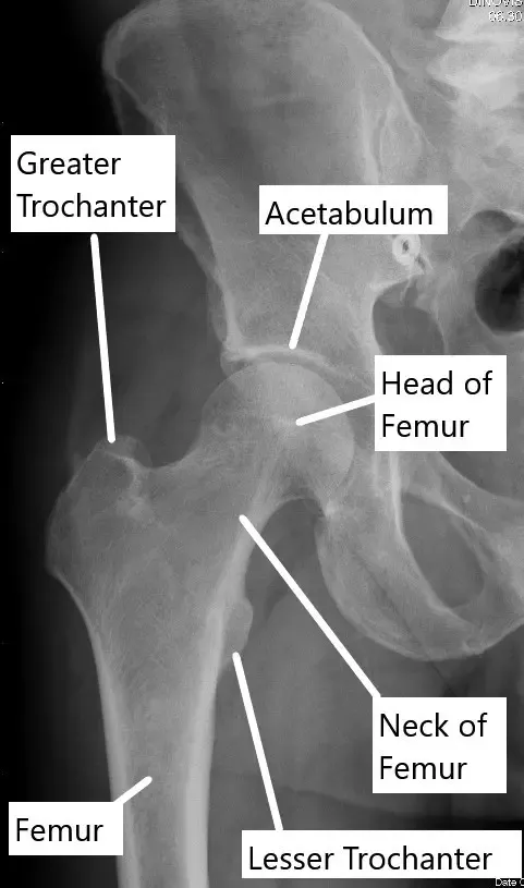
X-ray showing normal anatomy of the hip joint.
Why It Happens – Causes (Etiology and Pathophysiology)
The exact cause of transient osteoporosis is not fully understood, but it is believed to result from temporary disruption of the blood supply to the femoral head. This disruption can cause swelling and reduced blood flow, leading to a loss of bone density. Over time, the density of the femoral head decreases, resulting in osteolysis (bone breakdown). In pregnant women, hormonal changes may also play a role. The condition is typically self-limiting, meaning the bone will naturally recover over time, usually within 12 months.
How the Body Part Normally Works? (Relevant Anatomy)
The hip joint is a ball-and-socket joint where the femoral head (ball) fits into the acetabulum (socket) of the pelvis. The joint is stabilized by a capsule of ligaments and muscles, and the surfaces are lined with cartilage that allows smooth movement. In transient osteoporosis, the femoral head loses its density and becomes more prone to pain and injury, especially during weight-bearing activities.
What You Might Feel – Symptoms (Clinical Presentation)
The primary symptom of transient osteoporosis of the hip is pain in the groin, buttock, or side of the hip. The pain worsens with weight-bearing activities such as walking, climbing stairs, or standing for long periods. As the bone weakens, patients may develop a limp, avoiding pressure on the affected hip. In severe cases, high-impact activities could lead to a hip fracture due to the reduced strength of the femoral head.
How Doctors Find the Problem? (Diagnosis and Imaging)
Diagnosis is based on a combination of medical history, physical examination, and imaging studies. During the physical exam, doctors assess the hip’s range of motion and tenderness. Early X-rays may appear normal, but later imaging typically shows decreased bone density in the femoral head. MRI scans can detect bone edema (swelling) and loss of density at an earlier stage. A CT scan provides a detailed view of the hip bones. A nuclear bone scan can also identify changes in bone activity earlier than other methods.
Classification
Transient osteoporosis of the hip is generally not classified into subtypes but is recognized by the sudden, localized loss of bone density in the femoral head. It differs from other conditions like avascular necrosis (AVN), which involves progressive bone death and often requires more aggressive treatment.
Other Problems That Can Feel Similar (Differential Diagnosis)
Transient osteoporosis can be mistaken for generalized osteoporosis, which affects the bones throughout the body, particularly in postmenopausal women. It may also be confused with avascular necrosis (AVN), a condition where the bone dies due to a lack of blood supply. Unlike transient osteoporosis, AVN typically causes long-term damage to both hips and often requires surgical intervention.
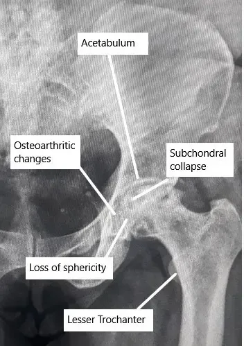
X-ray showing advanced AVN changes in the Hip.
Treatment Options
Non-Surgical Care – Most cases of transient osteoporosis resolve with nonsurgical management, which focuses on reducing pain and preventing further strain on the affected hip:
- Pain management with nonsteroidal anti-inflammatory drugs (NSAIDs) like ibuprofen or naproxen.
- Partial weight-bearing or using a cane to offload pressure from the joint.
- Physical therapy to maintain muscle strength and flexibility while avoiding strain on the hip.
Surgical Care – Surgery is rarely needed for transient osteoporosis, as the condition generally resolves on its own. However, in severe cases or when bone fractures occur, surgery may be considered to stabilize the joint.
Recovery and What to Expect After Treatment
Patients typically recover fully from transient osteoporosis within one to one and a half years. During recovery, pain usually subsides as the bone density returns to normal. The use of a cane or crutches during the healing period helps reduce strain on the affected hip. Most patients experience complete resolution of symptoms without long-term disability.
Possible Risks or Side Effects (Complications)
The main risk associated with transient osteoporosis is a potential fracture if the femoral head becomes too weak. This can occur if high-impact activities are performed before the bone has fully recovered. Otherwise, the condition is generally self-limiting, and there are few long-term complications once the bone density returns to normal.
Long-Term Outlook (Prognosis)
The long-term outlook for patients with transient osteoporosis of the hip is excellent. Most individuals recover fully, with no lasting joint deformities or chronic pain. Once the bone density is restored, the hip usually regains normal function.
For insurance and cost information, see our Insurance Information page.
Frequently Asked Questions (FAQ)
Q. Can transient osteoporosis of the hip cause permanent damage?
A. No, transient osteoporosis is typically a self-limiting condition, and the bone usually heals fully within a year.
Q. What are the main symptoms?
A. Pain in the groin, buttock, or side of the hip, especially with weight-bearing activities. The pain may worsen over time and can lead to limping.
Q. How is transient osteoporosis different from avascular necrosis?
A. Avascular necrosis causes bone death due to disrupted blood flow, typically leading to permanent joint damage. In contrast, transient osteoporosis is a temporary condition with a full recovery.
Summary and Takeaway
Transient osteoporosis of the hip is a rare condition that causes temporary bone density loss in the femoral head, leading to hip pain and limping. The condition is usually self-limiting and resolves within 12 months with nonsurgical treatment such as pain management and physical therapy. Full recovery is expected, and long-term complications are rare.
Clinical Insight & Recent Findings
Transient osteoporosis of the hip joint is a rare but temporary condition that causes hip pain and a reduction in bone density in the femoral head. It is typically self-limiting, with most patients experiencing full recovery within 12 months.
A recent case study underscores the difficulty of diagnosing ITOH, as its symptoms often resemble those of more serious conditions like avascular necrosis. The patient in the study underwent a thorough diagnostic process, including MRI, which revealed characteristic bone marrow edema. After a conservative treatment regimen, including partial weight-bearing and pain relief, the patient fully recovered.
This case highlights the importance of considering ITOH in patients with unexplained hip pain, especially when other serious conditions have been ruled out. (“Study on idiopathic transient osteoporosis of the hip – see PubMed.”)
Who Performs This Treatment? (Specialists and Team Involved)
Orthopedic specialists, particularly those focused on hip conditions, manage transient osteoporosis of the hip. Physical therapists assist with rehabilitation to improve mobility and strength. Radiologists provide essential imaging studies for diagnosis.
When to See a Specialist?
If you experience hip pain that doesn’t improve with rest, or if pain worsens during weight-bearing activities, see an orthopedic specialist for evaluation and diagnosis.
When to Go to the Emergency Room?
Go to the emergency room if you experience sudden severe pain, difficulty walking, or if your hip becomes swollen or bruised after an injury.
What Recovery Really Looks Like?
Recovery is typically gradual, with pain subsiding over months as the bone heals. Patients may use a cane to reduce pressure on the hip during recovery. Most people can return to normal activities after one to one and a half years without any long-term issues.
What Happens If You Ignore It?
Ignoring transient osteoporosis may lead to fractures if the bone becomes too weak, especially during high-impact activities. Early intervention and monitoring prevent this risk.
How to Prevent It?
Maintain bone health through proper nutrition, including calcium and vitamin D, and regular weight-bearing exercises. Avoid high-impact activities if you have hip pain, and follow your doctor’s recommendations for activity modification during recovery.
Nutrition and Bone or Joint Health
A diet rich in calcium, vitamin D, and protein supports bone strength and healing. Adequate hydration and a healthy weight also help reduce strain on the hip joint during recovery.
Activity and Lifestyle Modifications
Low-impact activities such as swimming or cycling are ideal during recovery. Stretching and strengthening exercises for the hip can improve flexibility and help prevent recurrence of hip issues.
Do you have more questions?
Is transient osteoporosis of the hip a common condition?
Transient osteoporosis of the hip is considered rare but can occur, particularly in certain demographics such as middle-aged men or pregnant women.
What causes transient osteoporosis of the hip?
The exact cause of transient osteoporosis of the hip is unknown, but it is believed to involve factors such as hormonal changes, reduced blood flow to the hip joint, and mechanical stress.
How is transient osteoporosis of the hip diagnosed?
Diagnosis typically involves a combination of clinical evaluation, imaging studies such as X-rays, MRI scans, and bone density tests, and ruling out other possible causes of hip pain.
What are the risk factors for developing transient osteoporosis of the hip?
Risk factors may include being male, middle-aged, or pregnant, as well as certain medical conditions such as hyperparathyroidism or corticosteroid use.
Can transient osteoporosis of the hip affect both hips simultaneously?
Yes, transient osteoporosis of the hip can affect both hips simultaneously, although it may initially present in one hip before involving the other.
Is transient osteoporosis of the hip associated with any complications or long-term effects?
In most cases, transient osteoporosis of the hip resolves on its own with appropriate management and does not lead to long-term complications. However, in some instances, it may progress to avascular necrosis of the hip joint.
What are the treatment options for transient osteoporosis of the hip?
Treatment typically focuses on pain management, reducing weight-bearing activities on the affected hip, physical therapy to maintain joint mobility and muscle strength, and, in some cases, medications to improve bone density.
How long does transient osteoporosis of the hip typically last?
Transient osteoporosis of the hip usually resolves spontaneously within a few months, although the duration can vary depending on individual factors and the severity of the condition.
Can transient osteoporosis of the hip recur after resolution?
While rare, transient osteoporosis of the hip can recur in some individuals, particularly if underlying risk factors persist or if there is inadequate management of the condition.
Are there any lifestyle modifications or precautions recommended for individuals with transient osteoporosis of the hip?
Yes, individuals with transient osteoporosis of the hip may be advised to avoid activities that exacerbate hip pain or increase stress on the joint, maintain a healthy weight, and ensure adequate intake of calcium and vitamin D to support bone health.
Can transient osteoporosis of the hip lead to permanent damage or disability?
In most cases, transient osteoporosis of the hip does not lead to permanent damage or disability if managed appropriately. However, in rare instances or severe cases, it may progress to avascular necrosis or other complications.
Are there any surgical interventions available for treating transient osteoporosis of the hip?
Surgical interventions are typically not necessary for transient osteoporosis of the hip, as the condition usually resolves with conservative management. However, in cases of severe pain or complications such as fracture, surgical options may be considered.
Can physical therapy or rehabilitation exercises worsen symptoms of transient osteoporosis of the hip?
Physical therapy and rehabilitation exercises are generally beneficial for maintaining joint mobility and muscle strength in transient osteoporosis of the hip, but it’s essential to avoid activities that exacerbate pain or discomfort.
Are there any alternative or complementary therapies that may help manage symptoms of transient osteoporosis of the hip?
While not a replacement for conventional medical treatment, some individuals may find relief from symptoms of transient osteoporosis of the hip through therapies such as acupuncture, chiropractic care, or herbal supplements. However, the effectiveness of these approaches varies, and consultation with a healthcare provider is recommended.
Can transient osteoporosis of the hip affect other joints in the body besides the hips?
While transient osteoporosis of the hip primarily affects the hip joints, it can theoretically affect other weight-bearing joints such as the knees or ankles, although this is less common.
Are there any specific precautions or considerations for pregnant women with transient osteoporosis of the hip?
Pregnant women with transient osteoporosis of the hip may require specialized management to ensure optimal pain relief and mobility while considering the safety of treatment options for both the mother and the fetus.
Can transient osteoporosis of the hip cause permanent changes in bone density or structure?
In most cases, transient osteoporosis of the hip does not cause permanent changes in bone density or structure, as it is a self-limiting condition that typically resolves without long-term effects.
How can I prevent transient osteoporosis of the hip from recurring in the future?
Preventing recurrence of transient osteoporosis of the hip may involve addressing underlying risk factors such as hormonal imbalances, avoiding excessive weight-bearing activities, maintaining a healthy lifestyle, and following any recommendations from healthcare providers for bone health maintenance.
Are there any specific medications or supplements that can help prevent or treat transient osteoporosis of the hip?
In some cases, medications such as bisphosphonates or calcitonin may be prescribed to improve bone density and reduce the risk of fracture in transient osteoporosis of the hip. Additionally, calcium and vitamin D supplements may be recommended to support bone health.
The content on this page has been authored, edited, or approved by the doctors below, and was last reviewed for accuracy on May 17, 2026.
