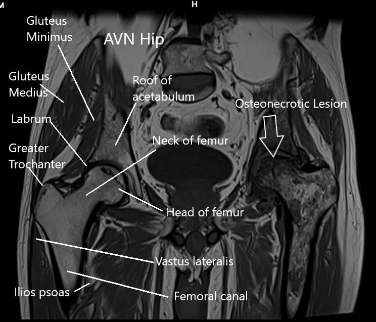
Long term alcohol intake is a common cause of avascular necrosis of the head of the femur. Chronic steroid intake and alcohol account for more than 60% of non-traumatic causes of avascular necrosis in the United States.
Avascular necrosis commonly affects the head of the femur due to its limited blood supply. The head of the femur (thighbone) forms the ball which fits into the cup formed by the acetabulum (pelvis). Any cause which may lead to disruption of the blood supply may cause avascular necrosis.
The disruption of the blood supply leads to the death of the bone cells in the head of the femur. The dead bone slowly collapses with time. The overlying joint cartilage collapses leading to an incongruous head of the femur.
X-ray of both the hip joints showing AVN of the left hip.
The loss of sphericity leads to arthritic changes in the cup and the ball. The joint becomes painful and stiff with loss of movement in various planes of motion.
The long term heavy drinking is a known cause of avascular necrosis AVN of the head of the femur. Most patients with alcohol-induced avascular necrosis usually belong to the ages of 50 years and above. The average duration of alcohol intake is usually 8 – 10 years with weekly consumption of 400 ml or more.

MRI of both the hip joints in the coronal section.
One of the effects of alcohol is hyperlipidemia. Hyperlipidemia is the excess of fatty cells in the body. The blood levels of fat in the form of triglycerides, low-density lipoproteins (LDL) and very-low-density lipoproteins (VLDL) increases.
The increased fat content of the blood causes blockage of the blood vessels. The head of the femur is especially susceptible to decreased blood flow and it may lead to aseptic necrosis/cell death.
Additionally, fat cells known as adipocytes increase in the head of the femur. This causes an increase in pressure inside the head of the femur. The increased pressure further decreases the blood flow inside the head femur.
Alcohol also acts as a direct cellular toxin causing damage to the bone cells. The affected cells undergo changes that ultimately lead to cell death/necrosis. The healthy bone gets replaced with dead and fibrotic tissues.
Usually, patients with a history of long-standing alcohol intake usually present when the avascular necrosis is in advanced stages. During the initial stages of avascular necrosis, the patients are generally asymptomatic. In subsequent stages, the patients may experience hip pain and stiffness.
The pain is usually dull aching in character located in the front of the hip. The patient may report exacerbation of pain with movements. The patients experience difficulty in getting up from sitting position, walking, navigating stairs, and bending.
The stiffness usually begins in one motion, eg patients may find it difficult to tie shoelaces. Gradually as the disease progresses, the stiffness limits movement in most planes of the movement.
The diagnosis of avascular necrosis is made by the physician through physical examination and imaging tests. An X-ray is helpful to look for the bone collapse in the head of the femur. Further, arthritic changes in the head of the femur and acetabulum can be evaluated with an X-ray.
The early stages of the disease process can be diagnosed with an MRI. MRI imaging is able to discern areas of decreased blood supply and necrosis. Bone scans are also useful in seeing areas of decreased blood supply. People who drink alcohol usually develop avascular necrosis of both the hips.
The management of avascular necrosis induced by alcohol depends upon the stage of the disease. In the early stages of the disease, both medical management and surgical management may be done. Medical management is done with lipid-lowering medications such as statin and blood thinners such as warfarin. Bisphosphonates may be used to decrease bone loss.
The medical management is accompanied by surgical procedures such as core decompression, vascularized grafts, or osteotomies. Core decompression involves the use of very small drills. These are used to drill holes in the head of the femur and may be accompanied with stem cell therapy. The goal in core decompression is to reduce pressure inside the head and increase repair.
Vascularized or non vascularized bone grafts may be inserted in the head of the femur. Osteotomies are bone cutting procedures aimed to rotate the diseased segment of the bone and allow healing. Even with these head preservation surgical procedures, a vast majority of patients end up with disease progression.
A total hip replacement usually is required in the majority of cases of avascular necrosis. The surgery involves the removal of the diseased parts (head of the femur) of the hip joint. Prosthetic metal and plastic parts are then inserted. The artificial hip joint recreates the movements of the natural hip.
The cup of the acetabulum is made of metal alloy or ceramic and is fixed with screws or press-fitted. A unique highly durable plastic is fixed in the cup for smooth gliding. A stem made of metal alloy is either press-fitted or fixed with bone cement inside the cavity of the thighbone. A prosthetic head made of metal alloy or ceramic is then fixed to the stem.
The improvement in the quality of life and the relief from pain is drastic in joint replacement surgery. The patients are able to get back to the lifestyle they enjoy quickly. With the advances in implant materials and surgical techniques, the surgeries today last for many years.

Dr. Suhirad Khokhar
My name is Dr. Suhirad Khokhar, and am an orthopaedic surgeon. I completed my MBBS (Bachelor of Medicine & Bachelor of Surgery) at Govt. Medical College, Patiala, India.
I specialize in musculoskeletal disorders and their management, and have personally approved of and written this content.
My profile page has all of my educational information, work experience, and all the pages on this site that I've contributed to.

