Anterior Cruciate Ligament (ACL) injuries are common among athletes and individuals involved in physical activities. When the ACL is torn, surgery is often needed to repair or reconstruct the ligament. However, there is a risk of graft failure post-surgery, which can lead to recurrent instability and pain in the knee. This article discusses the causes and symptoms of ACL surgery graft failure, providing insights in a simple and clear manner.
How Common It Is and Who Gets It? (Epidemiology)
ACL injuries are common in sports, particularly those involving running, jumping, and pivoting, such as basketball, soccer, and skiing. ACL reconstruction surgery is a frequent treatment, but graft failure is a potential complication. Graft failure is more likely to occur in active individuals, especially athletes who return to high-intensity activities too soon after surgery. The condition may also be more prevalent in individuals with poor rehabilitation compliance or those with anatomical issues affecting graft placement.
Why It Happens – Causes (Etiology and Pathophysiology)
Graft failure after ACL surgery can occur for several reasons:
- Re-injury: Athletes or active individuals who return to sports too soon may cause additional stress on the graft, leading to rupture.
- Surgical Technique: Poor graft placement or incorrect tunnel positioning can lead to graft failure. Mal-positioning of the graft can create abnormal stress, causing loosening or failure.
- Weak Graft Fixation: If the graft is not securely fixed to the bones at the shin or thigh, it can loosen or rupture.
- Immunological Rejection (Allograft): Graft rejection is more common with donor tissue (allografts), especially in younger patients, causing inflammation and failure.
- Excessive Rehabilitation: Over-aggressive rehabilitation can lead to graft loosening or failure due to overloading.
- Additional Injuries: Missed injuries, such as meniscal tears or posterolateral corner injuries, can contribute to instability and affect the success of the graft.
- Poor Healing: Inadequate graft healing, often due to systemic conditions like diabetes, can result in graft failure.
How the Body Part Normally Works? (Relevant Anatomy)
The ACL is a key ligament in the knee that helps stabilize the joint during movements such as running, jumping, and pivoting. It connects the thighbone (femur) to the shinbone (tibia), preventing the tibia from moving too far forward relative to the femur. This ligament is essential for maintaining knee stability and functionality during physical activity.
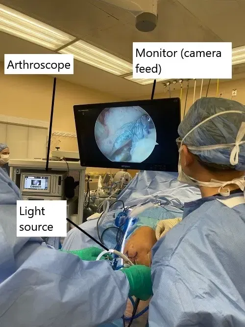
Intraoperative image of ACL reconstruction.
What You Might Feel – Symptoms (Clinical Presentation)
Recognizing the symptoms of ACL graft failure is crucial for timely intervention. Common symptoms include:
- Recurrent Instability: A feeling of the knee “giving way” or buckling, especially during physical activities or movements that stress the knee.
- Persistent Pain: Continuous or recurring pain, particularly during activities like bending, twisting, or squatting.
- Swelling: Swelling around the knee joint, which may worsen after physical activity or prolonged standing.
- Decreased Range of Motion: Difficulty fully extending or flexing the knee, leading to stiffness and discomfort.
- Feeling of Giving Way: A sensation of instability even during everyday activities like walking or climbing stairs.
- Decreased Functionality: A decrease in knee function, making simple tasks like walking or standing for long periods more challenging.
How Doctors Find the Problem? (Diagnosis and Imaging)
Diagnosis of ACL graft failure is based on a thorough physical examination and radiological tests. Physical tests, such as checking for knee instability or alignment, are performed. Imaging studies, including X-rays, MRI, and CT scans, may be used to assess the position of the graft, bone tunnel alignment, and any other potential issues like meniscal tears. MRI is particularly useful for visualizing soft tissue and graft status.
Classification
ACL graft failure is generally classified into early and late failure:
- Early failure (within the first 12 months) is usually due to non-traumatic causes, such as poor graft fixation or surgical errors.
- Late failure (after 12 months) is often due to re-injury or excessive physical stress on the graft, especially in athletes.
Other Problems That Can Feel Similar (Differential Diagnosis)
Conditions such as meniscal tears, cartilage damage, or ligament injuries (e.g., posterolateral corner injuries) may present symptoms similar to those of ACL graft failure. It is essential to differentiate these conditions from graft failure for proper treatment.
Treatment Options
Non-Surgical Care
In some cases, conservative management may help alleviate symptoms, including:
- Physical Therapy: To strengthen the muscles around the knee and improve stability.
- Bracing: A knee brace may provide additional support during daily activities or sports.
- Pain Management: NSAIDs or corticosteroid injections may help reduce pain and inflammation.
Surgical Care
If graft failure is confirmed, revision ACL surgery is usually required. The two primary options are:
- Single-Stage Revision: A single surgery to remove the failed graft and place a new one. This may involve using either an autograft or an allograft.
- Two-Stage Revision: A more complex approach that may be needed if bone grafting or additional interventions (e.g., osteotomy) are required. This may be used in cases with significant bone damage or mal-alignment.
Recovery and What to Expect After Treatment
Recovery from ACL revision surgery typically involves a rehabilitation program similar to the original ACL surgery. This includes:
- Initial Phase: Focus on reducing swelling and restoring knee motion.
- Strengthening Phase: Gradual strengthening of the quadriceps and hamstrings.
- Return to Activity: Once strength and stability are regained, patients can slowly return to normal activities, usually within 6 to 9 months for high-impact activities.
Possible Risks or Side Effects (Complications)
Complications of revision ACL surgery include:
- Infection: As with any surgery, infection is a risk.
- Nerve Injury: Nerves around the knee can be injured during surgery.
- Graft Failure: Even with revision surgery, there is a risk that the new graft may fail again.
- Scar Tissue: Excessive scarring may limit the knee’s range of motion or lead to further instability.
Long-Term Outlook (Prognosis)
With proper treatment, most patients can regain full functionality after revision ACL surgery. However, the long-term success of the graft depends on adherence to rehabilitation protocols, avoiding re-injury, and ensuring proper surgical technique. The prognosis for those who experience graft failure is generally good, although there may be some ongoing risks of re-injury, especially for athletes.

Instrument (co-ablation wand) used in knee arthroscopic surgery.
For insurance and cost information, see our Insurance Information page.
Frequently Asked Questions (FAQ)
Q. What causes ACL graft failure?
A. ACL graft failure can be caused by poor graft fixation, surgical errors, re-injury, or improper rehabilitation. Other factors such as immunological rejection (in the case of allografts) or missed injuries (e.g., meniscal tears) can contribute.
Q. How is ACL graft failure diagnosed?
A. ACL graft failure is diagnosed through a combination of physical examination, imaging studies (such as MRI and CT scans), and evaluation of the patient’s symptoms and history.
Q. Can ACL graft failure be prevented?
A. While not all cases of graft failure can be prevented, following rehabilitation guidelines, avoiding premature return to sports, and maintaining good knee health can help reduce the risk of graft failure.
Q. What is the recovery time for ACL revision surgery?
A. Recovery typically takes 6 to 9 months, with a gradual return to normal activities. Full recovery, especially for high-impact activities, may take up to a year.
Summary and Takeaway
ACL graft failure is a serious but manageable complication of ACL surgery. Early recognition of symptoms such as knee instability, persistent pain, and swelling can help prevent further damage and guide appropriate treatment. Revision surgery can often restore knee function, but it requires careful rehabilitation and adherence to recovery protocols to ensure long-term success.
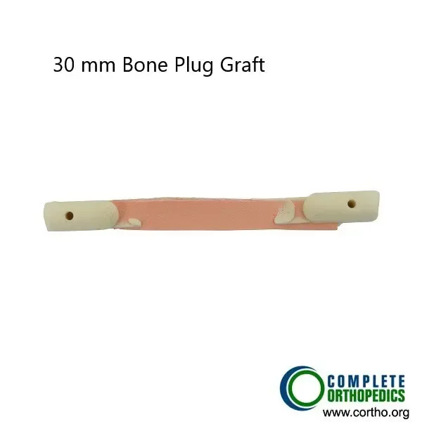
Bone plug graft used in ACL reconstruction.
Who Performs This Treatment? (Specialists and Team Involved)
ACL reconstruction and revision surgeries are typically performed by orthopedic surgeons, specifically those specializing in sports medicine or knee surgery. Physical therapists play an essential role in rehabilitation following surgery.
When to See a Specialist?
If you experience instability, pain, or swelling in the knee after ACL surgery, it’s important to consult with your surgeon or healthcare provider for a thorough evaluation.
When to Go to the Emergency Room?
You should seek emergency care if you experience sudden, severe pain, a noticeable shift in knee alignment, or signs of infection such as redness, warmth, or fever after surgery.
What Recovery Really Looks Like?
Recovery involves physical therapy and a gradual return to activities. Patients typically need to follow a rehabilitation program, with an emphasis on strengthening the knee and improving range of motion. Full recovery for high-impact sports may take up to 12 months.
What Happens If You Ignore It?
Ignoring symptoms of graft failure can lead to further damage to the knee joint, including degenerative changes and chronic instability. Timely intervention is critical to prevent permanent damage.
How to Prevent It?
Following rehabilitation protocols, avoiding premature return to sports, maintaining overall knee health, and communicating with your healthcare provider can reduce the risk of ACL graft failure.
Nutrition and Bone or Joint Health
Maintaining a balanced diet rich in nutrients like calcium and vitamin D is essential for bone and joint health, particularly after surgery. Regular exercise and maintaining a healthy weight also help reduce stress on the knee.
Activity and Lifestyle Modifications
Avoiding high-impact activities too soon after surgery, practicing proper biomechanics, and gradually returning to sports can help prevent ACL graft failure and improve long-term knee health.
For ACL procedure and cost information, please click here.
Do you have more questions?
How common is ACL surgery graft failure?
ACL surgery graft failure occurs in a small percentage of cases, estimated to be around 5% to 15%, depending on various factors such as patient age, activity level, and surgical technique.
Can ACL surgery graft failure occur immediately after surgery?
While it’s possible for graft failure to occur shortly after surgery due to factors such as graft tension or technical errors during the procedure, it’s more common for graft failure to occur months or even years later.
Are there different types of grafts used in ACL surgery, and do they affect the risk of graft failure?
Yes, there are different types of grafts used in ACL surgery, including autografts (such as the patellar tendon, hamstring tendon, or quadriceps tendon) and allografts (donor tissue). While the choice of graft may influence the risk of graft failure, the success of surgery depends on various factors beyond just the type of graft.
What are the potential complications of revision ACL surgery for graft failure?
Revision ACL surgery for graft failure carries similar risks as primary ACL surgery, including infection, stiffness, and persistent instability. Additionally, the presence of scar tissue from the previous surgery may complicate the revision procedure.
How long does it take to recover from revision ACL surgery?
Recovery from revision ACL surgery can take longer than primary ACL surgery, often requiring several months of rehabilitation and gradual return to activities.
Is ACL surgery graft failure more common in certain age groups?
ACL surgery graft failure can occur in patients of all age groups, but it may be more common in younger patients due to higher activity levels and potential for reinjury.
Are there specific factors that increase the risk of ACL surgery graft failure?
Yes, several factors can increase the risk of ACL surgery graft failure, including inadequate rehabilitation, premature return to sports, technical errors during surgery, and individual biological factors.
Can ACL surgery graft failure be prevented?
While not all cases of graft failure can be prevented, patients can take steps to reduce the risk by following rehabilitation guidelines, avoiding premature return to sports, and maintaining good knee health.
What should I do if I suspect graft failure after ACL surgery?
If you experience symptoms such as recurrent instability, persistent pain, or swelling after ACL surgery, it’s important to consult with your orthopedic surgeon for a thorough evaluation and appropriate management.
Are there alternative treatments for ACL injuries besides surgery?
In some cases, nonsurgical treatments such as physical therapy and activity modification may be recommended for ACL injuries, particularly for individuals with lower activity levels or specific anatomical factors.
How long does it typically take for a graft to heal after ACL surgery?
The time it takes for a graft to heal after ACL surgery can vary depending on factors such as graft type, patient age, and adherence to rehabilitation protocols. Generally, it takes several months for the graft to fully integrate with surrounding tissues.
Can ACL surgery graft failure lead to long-term complications?
Yes, ACL surgery graft failure can lead to long-term complications such as persistent instability, recurrent injuries, and accelerated joint degeneration (osteoarthritis) if left untreated.
Are there any dietary or lifestyle changes that can help promote graft healing after ACL surgery?
While maintaining a balanced diet and healthy lifestyle can support overall healing and recovery after ACL surgery, there are no specific dietary or lifestyle changes proven to directly promote graft healing.
Is it possible to return to sports or physical activities after experiencing graft failure?
Yes, with appropriate treatment and rehabilitation, many patients can return to sports or physical activities after experiencing graft failure. However, the timing and feasibility of return to activities depend on individual factors and the severity of the graft failure.
Can ACL surgery graft failure occur in both knees?
Yes, ACL surgery graft failure can occur in one or both knees, particularly in individuals who participate in high-impact sports or activities that place repeated stress on the knees.
Are there any warning signs that indicate an increased risk of ACL surgery graft failure?
While there are no definitive warning signs for ACL surgery graft failure, patients should be vigilant for symptoms such as recurrent instability, persistent pain, or swelling after surgery, as these may indicate a potential problem with the graft.
How can I prevent reinjury after ACL surgery?
To prevent reinjury after ACL surgery, it’s important to follow rehabilitation guidelines, gradually increase activity levels, and use appropriate protective equipment (such as braces) during sports or high-risk activities.
Are there any specific exercises or activities I should avoid after ACL surgery to reduce the risk of graft failure?
Patients should avoid high-impact activities, sudden changes in direction, and heavy lifting during the initial stages of rehabilitation to reduce the risk of graft failure. Your physical therapist can provide guidance on safe exercises and activities.
Can ACL surgery graft failure be detected on imaging studies such as X-rays or MRI?
Imaging studies such as MRI can help assess the integrity of the reconstructed ACL graft and identify signs of graft failure, such as graft laxity or abnormal signal intensity.
Is it normal to experience swelling and discomfort in the knee after ACL surgery, even if the graft is intact?
Yes, swelling and discomfort are common after ACL surgery due to the trauma of the procedure and the body’s healing response. However, if symptoms persist or worsen over time, further evaluation may be necessary to rule out graft failure or other complications.
Are there any medications that can help prevent graft failure after ACL surgery?
There are no specific medications proven to prevent graft failure after ACL surgery. However, your surgeon may prescribe pain medications or anti-inflammatory drugs to manage symptoms during the recovery period.
How soon can I return to driving after ACL surgery, and does it increase the risk of graft failure?
The timing of return to driving after ACL surgery depends on factors such as the type of surgery, the use of pain medications, and individual comfort level. Driving typically becomes feasible once patients regain sufficient strength and range of motion in the knee, but it’s important to consult with your surgeon for personalized guidance. Driving itself does not inherently increase the risk of graft failure, but patients should avoid driving if they experience significant pain, stiffness, or limitations in mobility that could affect their ability to operate a vehicle safely.
Are there any psychological effects associated with experiencing graft failure after ACL surgery?
Experiencing graft failure after ACL surgery can be emotionally challenging for patients, as it may necessitate additional treatment and delay return to sports or activities. Patients may experience feelings of frustration, disappointment, or anxiety about reinjury. It’s important for patients to communicate openly with their healthcare providers and seek support from family, friends, or mental health professionals if needed to cope with these emotions and maintain a positive outlook on their recovery.
The content on this page has been authored, edited, or approved by the doctors below, and was last reviewed for accuracy on May 15, 2026.
