A 47-year-old female presented with right foot pain following an acute injury. She reported direct trauma to the foot at work. Initially, the pain was mild but progressively worsened, leading to difficulty bearing weight. She described localized swelling and tenderness along the first and second metatarsals. The patient denied any numbness, tingling, or systemic symptoms such as fever or chills.
She has a history of high physical activity levels due to her occupation, requiring prolonged standing and walking. She denied prior foot injuries or orthopedic conditions. However, she admitted to continuing to walk on the injured foot for several days before seeking medical attention, potentially exacerbating the fractures.
Examination and Diagnosis
Upon physical examination, there was moderate swelling and tenderness over the dorsal aspect of the right foot. Bruising was noted over the first and second metatarsals. The patient exhibited a limited active and passive range of motion due to pain. Capillary refill was intact, and there were no signs of neurovascular compromise. The contralateral extremity demonstrated a normal range of motion, stability, and strength.
Radiographic Findings
Displaced fractures of the first and second metatarsals
Disruption of the metatarsophalangeal joint alignment
No acute dislocations or additional fractures
Given the severity and risk of malunion, open reduction and internal fixation (ORIF) was recommended to restore alignment and stability.
Preoperative Imaging
An X-ray of the right foot confirmed fractures of the first and second metatarsals.
Surgical Management
After obtaining informed consent, the patient underwent ORIF of the first and second metatarsals under regional anesthesia. The procedure was performed as follows:
A dorsal incision was made over the metatarsal shafts, with subcutaneous dissection down to the fracture sites.
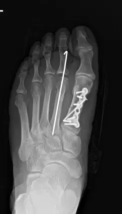
The fractures were reduced and temporarily stabilized with K-wires.
Definitive fixation was achieved using titanium plates and screws.
Fluoroscopic imaging confirmed anatomic alignment and stable fixation.
The surgical site was irrigated, and closure was performed in layers using absorbable sutures for deep tissues and non-absorbable sutures for the skin.
A well-padded dressing was applied, and the foot was immobilized in a posterior splint.
Postoperative Course
The patient was monitored in the recovery unit and reported well-controlled pain. She was discharged with a pneumatic walking boot and instructed to remain non-weight-bearing on the affected foot. Pain management included hydromorphone 2 mg, naproxen 500 mg, and acetaminophen 500 mg. Strict postoperative care instructions were provided, including elevation, icing, and avoidance of premature weight-bearing.
First Follow-Up (3 Weeks Post-Op)
At the initial follow-up, the patient reported persistent pain but admitted to noncompliance with weight-bearing restrictions. She had removed her own splint and sutures at home, against medical advice, and expressed a strong desire to remove the second metatarsal pin early.
Physical examination revealed mild swelling and tenderness but no erythema or signs of infection. Repeat X-rays showed early fracture healing but incomplete consolidation of the second metatarsal.
Given the risk of delayed healing, strict non-weight-bearing instructions were reinforced, and the patient was advised to continue using the pneumatic boot. Physical therapy was initiated to maintain foot and toe mobility and prevent stiffness.
Second Follow-Up (6 Weeks Post-Op)
At six weeks postoperatively, the patient continued to struggle with compliance due to work-related challenges. However, she reported a significant reduction in pain. Examination showed continued healing, with no erythema, drainage, or infection. Given the patient’s lifestyle constraints, she was transitioned to partial weight-bearing as tolerated, with continued use of the pneumatic boot.
Follow-up X-rays demonstrated progressive healing of the fractures.
Final Follow-Up (3 Months Post-Op)
At three months, the patient demonstrated substantial improvement. She reported minimal pain, full weight-bearing without discomfort, and no longer required pain medications. Repeat X-rays confirmed excellent callus formation and maintained alignment of the metatarsals.
At this stage, she was cleared for full weight-bearing without restrictions and advised to continue strengthening exercises. She successfully resumed daily activities, including work, without limitations.
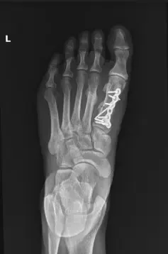
Postoperative internal fixation with plate and screws of a fracture of the first metatarsal.
Rehabilitation Plan and Long-Term Outcome
The patient was prescribed a structured rehabilitation program, including:
Strengthening exercises to improve foot stability and mobility.
Gradual return to full weight-bearing activities over the next 4-6 weeks.
Monitoring for potential complications, such as nonunion or persistent pain.
Despite early non-adherence, the patient’s commitment to physical therapy in the later stages contributed to her successful recovery. She continues to follow up as needed for long-term monitoring.
Conclusion
This case highlights the importance of postoperative compliance in fracture healing. Despite early non-adherence, structured treatment, patient education, and progressive rehabilitation led to a successful recovery. Long-term monitoring will ensure continued stability and function.
Disclaimer: Patient’s name, age, sex, dates, and events have been modified to protect patient privacy.
