

Knee instability, often described as a feeling of the knee “giving away” or buckling, can significantly impact daily life. This condition is commonly associated with pain, swelling, and difficulty with activities that require knee stability. Whether caused by ligament tears, meniscus injuries, or other underlying issues, understanding the causes and management strategies for knee instability is crucial for recovery and maintaining a good quality of life.
How Common It Is and Who Gets It? (Epidemiology)
Knee instability affects people of all ages but is particularly prevalent in athletes and individuals who engage in high-impact or twisting sports, such as football, soccer, and skiing. It is also common in older adults with degenerative knee conditions. Injuries to the knee ligaments, especially the anterior cruciate ligament (ACL), are a leading cause of knee instability. This condition often leads to functional limitations, including difficulty with walking, climbing stairs, or participating in recreational activities.
Why It Happens – Causes (Etiology and Pathophysiology)
Knee instability often results from injuries to the ligaments, meniscus, or other stabilizing structures within the knee. The most common causes include:
- ACL injuries: Typically caused by sudden twisting or pivoting movements.
- PCL injuries: Occur from direct force to the front of the knee, often seen in motor vehicle accidents or sports.
- Medial Collateral Ligament (MCL) injuries: Caused by a force to the outer side of the knee, often in contact sports.
- Meniscus tears: These can occur with rotational movements of the knee, leading to instability.
These injuries result in the loss of stability and function in the knee joint, contributing to a feeling of buckling.
How the Body Part Normally Works? (Relevant Anatomy)
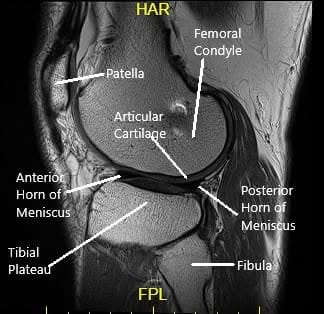
The knee joint is a hinge joint composed of the thigh bone (femur), shin bone (tibia), fibula, and kneecap (patella). The ligaments, cartilage, and menisci work together to provide stability and allow smooth movement.
- Articular cartilage cushions and protects the bones.
- Menisci serve as shock absorbers between the femur and tibia.
- Ligaments, including the ACL, PCL, MCL, and LCL, stabilize the knee by preventing excessive movement.
- Synovium produces synovial fluid, which lubricates the joint to ensure smooth motion.
When these structures are damaged, knee stability is compromised, leading to instability and an increased risk of injury.

Ligaments
The ligaments of the knee along with the meniscus provide stability to the knee. Tears or rupture of the ligaments and menisci are the most common cause of knee buckling.
Anterior Cruciate Ligament (ACL)
The ACL runs from the front and middle of the tibia to the outer and backward side of the femur. The ligament provides stability mainly in the front and back of the knee. The ligament also stabilizes the rotatory movement of the knee.
Posterior Cruciate Ligament (PCL)
The PCL is present behind the knee joint. Along with ACL, it provides stability in the front and back of the knee. Similar to ACL, the PCL also provides rotational stability.
Medial & Lateral Collateral Ligaments
The medial collateral ligament provides stability on the inner side of the knee. Similarly, the lateral collateral ligament provides stability on the outer side of the knee.
What You Might Feel – Symptoms (Clinical Presentation)
The primary symptom of knee instability is the sensation of the knee “giving away” or buckling, especially during activities that involve twisting or turning. Other symptoms include:
- Knee pain
- Swelling
- Limited range of motion
- Weakness or difficulty bearing weight on the affected leg
These symptoms are typically worse during physical activities that place strain on the knee joint, such as running, climbing stairs, or even walking on uneven surfaces.
How Doctors Find the Problem? (Diagnosis and Imaging)
Knee instability is diagnosed through a combination of physical examination and imaging tests.
- Physical examination: The orthopedic surgeon assesses the knee’s stability by performing special maneuvers that stress the ligaments and examine the range of motion.
- X-rays: These provide a view of the bones and can help identify fractures or signs of arthritis.
- MRI: An MRI is essential for detecting soft tissue injuries, such as tears in ligaments, the meniscus, or cartilage.
- Arthroscopy: Considered the gold standard, this procedure involves inserting a small camera into the knee joint to directly visualize damage to the ligaments and cartilage.
Classification
Knee instability can be classified based on the location and severity of the ligament damage:
- Anterior Instability: Due to damage to the ACL, lateral capsular, or medial capsular ligaments.
- Posterior Instability: Due to damage to the PCL or arcuate complex.
- Medial Instability: Caused by injuries to the MCL, medial capsular ligament, and occasionally the PCL.
- Lateral Instability: Caused by injuries to the lateral collateral ligament, lateral capsular, or iliotibial band (ITB).
Other Problems That Can Feel Similar (Differential Diagnosis)
Other conditions that can mimic knee instability include:
- Patellar dislocation or subluxation
- Meniscus tears: May cause pain and a feeling of instability, particularly with twisting motions.
- Osteoarthritis: Can cause pain, swelling, and mechanical symptoms that resemble instability.
- Referred pain: Conditions such as hip or lower back issues can sometimes present as knee instability.
Treatment Options
Non-Surgical Care
For mild to moderate knee instability, non-surgical treatments may be effective, including:
- Bracing: To provide support and prevent excessive movement of the knee.
- Rest and ice: To reduce swelling and inflammation.
- Physical therapy: Strengthening the muscles around the knee, particularly the quadriceps, can help stabilize the joint.
- Anti-inflammatory medications: To manage pain and swelling.
Surgical Care
In more severe cases, surgery may be required to repair torn ligaments or other damaged structures. The most common surgical approach is arthroscopic surgery, in which small incisions are made, and a camera is used to guide the repair of the damaged ligaments or meniscus. Rehabilitation following surgery is crucial for regaining strength and mobility.
Recovery and What to Expect After Treatment
Recovery depends on the severity of the injury and the type of treatment. After non-surgical treatment, patients typically experience gradual improvement in knee stability, with most returning to normal activities within 6-8 weeks.
For surgical cases, recovery can take several months, with a structured rehabilitation program that includes physical therapy and gradual reintroduction to physical activities. Full recovery may take up to 6-12 months, depending on the extent of the injury and the success of the surgery.
Possible Risks or Side Effects (Complications)
Complications from knee instability treatment can include:
- Infection (especially after surgery)
- Blood clots: Especially after surgery, patients may be at risk of deep vein thrombosis (DVT).
- Re-injury: The knee may remain vulnerable to further injury if rehabilitation is not followed carefully.
- Chronic instability: In some cases, even after treatment, patients may continue to experience instability if the knee does not heal correctly.
Long-Term Outlook (Prognosis)
The prognosis for knee instability varies. In many cases, proper treatment and rehabilitation can restore knee stability and function. However, untreated or poorly managed knee instability can lead to further damage to the knee joint, including arthritis and ongoing instability.
For insurance and cost information, see our Insurance Information page.
Frequently Asked Questions (FAQ)
Q. Can knee instability be prevented?
A. While it is not always preventable, strengthening the muscles around the knee and using proper techniques during sports can reduce the risk of injury.
Q. How long will it take to recover from knee instability?
A. Recovery time depends on the severity of the injury and the treatment method. Non-surgical treatments may take a few weeks, while surgical recovery can take 6-12 months.
Q. Is surgery necessary for knee instability?
A. Surgery may be necessary if conservative treatments such as physical therapy and bracing do not provide sufficient relief or if there are significant ligament tears.
Summary and Takeaway
Knee instability is a common and often disabling condition that can impact daily activities. Early diagnosis and appropriate treatment, whether conservative or surgical, are key to managing the condition and improving outcomes. With proper care, most patients can regain stability and resume their regular activities.
Clinical Insight & Recent Findings
A recent study explored the outcomes of in-situ mosaicplasty fixation for treating unstable osteochondritis dissecans (OCD) of the knee in skeletally mature patients. This technique, which involves using autologous osteochondral plugs to stabilize the lesion, demonstrated high healing rates, with MRI scans showing complete healing in 12 out of 13 cases within six months.
The study highlighted that in-situ mosaicplasty offers both mechanical stability and biological augmentation, making it an ideal first-line surgical option for unstable OCD.
Additionally, it showed that the procedure is minimally invasive, with excellent patient-reported outcomes and minimal complications, further validating its use in managing unstable knee conditions. (“Study of in-situ mosaicplasty fixation for unstable knee OCD – See PubMed.“)
Who Performs This Treatment? (Specialists and Team Involved)
Orthopedic surgeons specializing in knee injuries typically perform surgery for knee instability. Physical therapists play a critical role in rehabilitation, while other specialists, such as pain management doctors, may assist in non-surgical management.
When to See a Specialist?
You should see a specialist if you experience knee instability that does not improve with rest or physical therapy, if you have significant knee pain or swelling, or if you are unable to bear weight on the affected leg.
When to Go to the Emergency Room?
Seek emergency care if you experience sudden, severe pain, inability to move the knee, or signs of a blood clot (e.g., swelling, redness, or warmth in the leg). These could indicate serious complications requiring immediate attention.
What Recovery Really Looks Like?
Recovery from knee instability varies depending on the severity of the injury and the chosen treatment. With appropriate care, most patients can return to their normal activities, though full recovery from surgery may take several months.
What Happens If You Ignore It?
Ignoring knee instability can lead to further damage to the knee, including arthritis, chronic pain, and increased risk of future injury. Early intervention is crucial for preventing long-term complications.
How to Prevent It?
Prevention includes strengthening the muscles around the knee, using proper form during physical activities, and wearing supportive gear, especially for athletes or individuals with a history of knee injuries.
Nutrition and Bone or Joint Health
A healthy diet rich in calcium, vitamin D, and protein is essential for maintaining strong bones and joints. Staying hydrated and maintaining a healthy weight can also reduce stress on the knee joint, preventing further instability.
Activity and Lifestyle Modifications
Patients with knee instability should focus on low-impact activities such as swimming or cycling while avoiding high-impact sports or movements that could exacerbate the condition. A physical therapist can recommend specific exercises to improve knee strength and stability.
Do you have more questions?
What exactly causes the knee to become unstable?
Knee instability is commonly caused by damage to ligaments such as the ACL, degenerative changes from osteoarthritis, or weakness in the muscles around the knee. It can also result from acute injuries or chronic wear and tear.
Are there specific exercises to prevent knee instability?
Yes, exercises focusing on strengthening the quadriceps, hamstrings, and calf muscles can help stabilize the knee. Balance exercises and core strengthening are also beneficial.
How is knee instability diagnosed?
Diagnosis typically involves a physical examination, patient history, and imaging tests like MRI or X-rays to assess ligament damage and joint status.
Can knee instability lead to other knee problems?
Yes, it can lead to increased wear and tear in the knee joint, exacerbate conditions like osteoarthritis, and increase the risk of falls and other injuries.
What are the treatment options for severe knee instability?
Severe instability might require surgical interventions such as ligament reconstruction or knee replacement, depending on the underlying cause.
How effective are knee braces in managing instability?
Knee braces can be very effective in providing support and stability, especially during activities that put stress on the knee.
What is the role of physical therapy in treating knee instability?
Physical therapy is crucial for strengthening the muscles around the knee, improving flexibility, and teaching stabilizing techniques to protect the joint.
Can knee instability be completely cured?
While some causes of instability can be effectively treated, chronic conditions like osteoarthritis may require ongoing management.
Is knee instability common in athletes?
Yes, athletes, particularly those involved in high-impact sports or activities that involve rapid direction changes, are at higher risk of developing knee instability.
What lifestyle changes can help manage knee instability?
Maintaining a healthy weight, avoiding high-impact activities, and regular knee-strengthening exercises can help manage symptoms.
How long does it take to recover from a procedure to correct knee instability?
Recovery times can vary widely based on the specific procedure, ranging from a few weeks to several months.
Are there age-specific concerns regarding knee instability?
Older adults may experience more pronounced effects due to muscle weakness and degenerative changes, while younger individuals may suffer from instability primarily due to injuries.
Can knee instability affect balance and coordination?
Yes, instability can significantly impact balance and coordination, increasing the risk of falls and affecting the ability to perform daily activities.
What are the signs that knee instability is worsening?
Increased frequency of knee giving way, heightened pain, swelling, and reduced mobility are signs that instability may be worsening.
Are there non-surgical treatments that can be effective?
Besides physical therapy and braces, treatments like corticosteroid injections, NSAIDs, and lifestyle modifications can be effective non-surgical options.
What is ACL reconstruction?
ACL reconstruction is a surgical procedure used to replace a torn anterior cruciate ligament, a common cause of knee instability.
How does obesity affect knee instability?
Obesity increases stress on the knee joints, exacerbating instability and associated symptoms like pain and reduced function.
Can knee instability be a sign of more serious health issues?
While it often relates to local issues within the knee, severe or unexplained instability should be evaluated to rule out other health problems.
What are the risks of surgery for knee instability?
Risks include infection, nerve damage, blood clots, and the potential for continued instability or pain.
How do I know if my knee instability is due to osteoarthritis?
A diagnosis typically involves evaluating symptoms like joint stiffness, pain during activity, and reviewing imaging studies.
What advancements are being made in treating knee instability?
Advances include new surgical techniques, better diagnostic tools, and developments in regenerative medicine like stem cell therapy.
Is swimming good for knee instability?
Yes, swimming is an excellent low-impact exercise that can strengthen the muscles around the knee without putting excessive stress on the joint.
How often should I perform stability exercises for my knee?
Frequency can vary based on individual needs, but generally, stability exercises should be performed 2-3 times per week, gradually increasing intensity and complexity under professional guidance.
What dietary considerations can help with knee health and stability?
A diet rich in anti-inflammatory foods such as omega-3 fatty acids, antioxidants, and adequate hydration can support joint health and potentially reduce symptoms related to knee instability.
The content on this page has been authored, edited, or approved by the doctors below, and was last reviewed for accuracy on May 25, 2026.